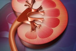
An imaging technology developed by the Mayo Clinic in Rochester, MN, is proving highly accurate in detecting liver fibrosis and may help eliminate the need for liver biopsies.
The technology, called magnetic resonance elastography (MRE), utilizes conventional 1.5-tesla MRI equipment and hardware to generate mechanical waves through the liver, which are then detected by a special pulse sequence designed by Mayo for the technique. The noninvasive procedure takes seconds to conduct, and focuses on calculating the stiffness of the liver, said Dr. Jayant Talwalkar, a hepatologist and associate professor of medicine at the Mayo Clinic.
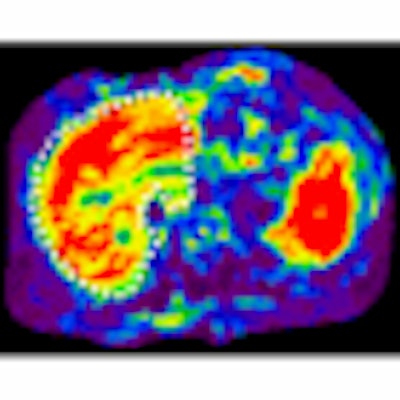
Elastogram colors
MRE produces color-coded images, or elastograms, that indicate how internal organs, muscles, and tissues would feel to the touch. The colors range from red, designating the stiffest areas, to purple, indicating the softest.
MRE research began at the Mayo Clinic about 10 years ago. "Initial experiments with healthy volunteers and a small group of patients with liver disease demonstrated the feasibility of using this test to measure hepatic fibrosis," said Talwalkar, who joined the effort about two years ago. "The initial study also gave us estimates of test performance, and we were pleasantly surprised that the initial study gave us an indication that the test would be quite accurate."
Mayo researchers last week released details of their latest MRE study at the annual gathering of the American Association for the Study of Liver Diseases (AASLD) in San Francisco.
The study included 113 patients who had undergone liver biopsy in the year preceding the study. The patients' liver diseases included nonalcoholic and alcoholic fatty liver disease, hepatitis C, hepatitis B, autoimmune hepatitis, primary biliary cirrhosis, and primary sclerosing cholangitis. Patients ranged in age from 19 to 78, and their body weight ranged from normal to severely obese.
Highly accurate detection
The analysis determined that MRE was highly accurate in detecting moderate-to-severe hepatic fibrosis in all ages, types of liver disease, and patient body sizes. In addition, the detection of cirrhosis by MRE, when compared to liver biopsy results, achieved an accuracy rate of 88%. Patients with nonalcoholic fatty liver disease and no significant inflammation or fibrosis were identified with 97%.
Previous MRE studies at Mayo also have shown the technology to be beneficial when comparing chronic liver disease patients with healthy volunteers. "Again we saw that MRE could accurately identify patients with severe fibrosis and distinguish them from patients with milder degrees of fibrosis," Talwalkar said.
 |
| The red areas of the elastogram show that liver is abnormally stiff, indicating presence of fibrosis. Image courtesy of the Mayo Clinic, Rochester, MN. |
Prior to MRE, Talwalkar said physicians were hampered to a degree by conventional tests -- liver biochemistries and imaging, such as ultrasound, CT, or MRI -- which were not sensitive enough to determine whether new drugs were having positive, negative, or negligible effects on patients. To determine if a patient had liver fibrosis, the gold standard generally was liver biopsy.
Talwalkar estimates that "at least half of the patients we see, if not more, would be spared [liver biopsy], because a number of patients have mild disease or no evidence of fibrosis. A small proportion of those patients have advanced fibrosis. As we incorporate the test more into clinical practice, we will get a better idea of exactly how many patients can avoid the need for a biopsy."
Expanding MRE
Mayo's other clinics in Jacksonville, FL, and Scottsdale, AZ, are starting to use the MRE technique, while the Mayo Clinic in Rochester incorporates more MRE into standard clinical practice.
"It has helped in our decision-making whether to send a patient for a biopsy," Talwalkar added. "Many times these patients are not significantly ill from their liver disease. They are at stages where liver function is still normal, but we believe that fibrosis is the precursor to cirrhosis. If we can identify patients with fibrosis and there is evidence this fibrosis is progressing, this is the time when existing and novel therapies can be applied to patients to halt disease progression."
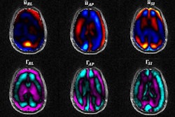
Research is under way to study how MRE might aid in the diagnosis of Alzheimer's disease and some cancers.
By Wayne Forrest
AuntMinnie.com staff writer
November 19, 2008
Related Reading
Magnetic resonance elastography identifies, quantifies myofascial taut bands, December 12, 2007
Scan painlessly pinpoints muscle stiffness, November 21, 2003
Copyright © 2008 AuntMinnie.com