NEW ORLEANS - Two images will share Image of the Year honors at this week's annual SNM conference, with Dr. Henry Wagner bestowing the award on images that demonstrate the versatility of hybrid imaging with different modalities.
Wagner, former SNM president and professor at Johns Hopkins University Bloomberg School of Public Health in Baltimore, unveiled the award-winning submissions on Monday at the SNM meeting.
"The four pillars of nuclear medicine and now molecular imaging are PET, SPECT, CT, and MRI," Wagner said. "These two papers employed all four pillars." The second reason for the selection was that both images "illustrate that molecular imaging extends from head to toe."
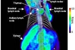
The first image, submitted by Policlinico S. Orsola-Universita di Bologna in Italy, uses PET/CT to reveal a relapse of neuroendocrine cancer. The study of a 33-year-old male patient was conducted with a radiotracer that revealed a suspicious lesion in the left ear, as well as the spread of cancer to the area of the pituitary gland and a lymph node in the neck.

The patient was referred for restaging after having received surgery a few months earlier.
Wagner said that the image is an excellent example of the advance in the "aesthetic characteristics" of PET/CT images.
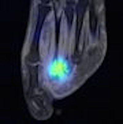
The second award-winning entry, from the Mount Sinai School of Medicine in New York City, shows the potential of extremity imaging with SPECT/MRI. The image is of a 24-year-old female patient known to have an infection in her foot. The issue was whether the patient had osteomyelitis.

"One of the problems is that most of the devices that are used for imaging are based on the use of photomultiplier tubes," Wagner said. "The blending of photomultiplier tubes in association with MRI has caused difficulties."
Several SNM papers have described the use of semiconductor detectors, but Wagner described them as "expensive imaging devices that are still in development." Still, the systems do not have photomultiplier tubes and "may be more applicable to hybrid fusing of the images."
In the Mount Sinai image, the SPECT study with labeled white blood cells was acquired on a separate gamma camera and fused with the images from an MRI scanner by computer superimposition to create the Image of the Year. Doctors concluded from the image that the patient had both an infection and osteomyelitis.
By Wayne Forrest
AuntMinnie.com staff writer
June 16, 2008
Related Reading
Nuclear medicine must overcome obstacles to flourish in the future, June 15, 2008
SNM explores feasibility of U.S. medical isotope source, May 22, 2008
Canadian government to stop development of Maple reactors, May 16, 2008
New U.S. budget funds nuclear medicine programs, January 14, 2008
Medical imaging averts cuts in new U.S. budget, December 20, 2007
Copyright © 2008 AuntMinnie.com