A pair of studies in this month's Radiology examines cardiac MRI's ability to predict adverse outcomes in patients with evidence of a previous myocardial infarction.
In the first study, researchers from Sweden examined patients with clinically unrecognized myocardial infarction (UMI) depicted at cardiac MRI to determine whether MI scars represented early damage from atherosclerosis, or perhaps a different pathogenesis. The presence of UMI has been shown to be a strong predictor of major adverse events.
In the second report, a team from Germany and California examined patients with previous acute myocardial infarction to determine whether infarct size could be used to predict left ventricular modeling, a strong prognostic factor indicating an elevated risk of future adverse events.
Unrecognized myocardial infarction
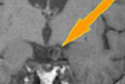
Late-enhancement MRI shows myocardial infarction (MI) and other myocardial scars as hyperintense regions, depicting both known and clinically unrecognized MI more accurately than electrocardiography, noted study authors Dr. Charlotte Barbier; Dr. Tomas Bjerner, Ph.D.; and colleagues from Uppsala University Hospital in Uppsala, Sweden (Radiology, October 2007, Vol. 245:1, pp. 103-110).
But while UMIs detected at electrocardiography are known to share similar risk factor profiles as recognized MIs (RMIs), it is unknown whether UMIs seen at MRI present a similarly high risk of future adverse events. Also unknown is whether these UMIs arise from early atherosclerosis or some other disease process.
Thus, the authors sought to investigate whether clinically unrecognized MIs detected at MRI have an atherosclerotic pathogenesis similar to that of recognized MIs.
The study examined a sample of 248 randomly chosen subjects (123 women, 125 men) who had undergone cardiac MRI as part of a large community-based screening program. "Subjects who had myocardial infarction scars at late-enhancement MR imaging on the original scans were classified as having RMI (n = 11) (those with a diagnosis of MI in the screening initiative) or UMI (n = 49) (those without a diagnosis of MI in the screening initiative)," Barbier and colleagues wrote.
For the present study, the team assessed Framingham risk scores for each participant with the aid of a questionnaire, blood pressure tests, and venous blood sampling to measure glucose, cholesterol, and C-reactive protein.
And to determine whether atherosclerosis was the cause of the scars, patients were rescanned with gadolinium-enhanced whole-body MR angiography (MRA), and late-enhancement cardiac scans a mean 16 months after the original cardiac MR scan.
MRA performed on a 1.5-tesla Gyroscan Intera system (Philips Medical Systems, Best, Netherlands) used a quadrature coil for MRA and a cardiac coil for cardiac imaging. Imaging included the aorta and the carotid, renal, and lower limb arteries to the ankle but not the coronary arteries, the authors wrote.
Whole-body images were acquired using a 3D radiofrequency spoiled T1-weighted gradient-echo sequence following administration of 40 mL of gadodiamide (Omniscan, GE Healthcare, Chalfont St. Giles, U.K.). Then cardiac late-enhancement images were acquired using a 3D inversion-recovery gradient-echo sequence that covered the heart in short- and long-axis views.
The presence of 50% or greater luminal narrowing in any vessel at whole-body MRA was considered to represent significant atherosclerosis. Ultrasound was used to measure the intima-media thickness of the common carotid artery in 239 of 248 subjects.
According to the results, the prevalence of significant atherosclerosis at whole-body MRA, intima-media thickness, C-reactive protein level, and Framingham risk score did not differ significantly between the group without MI scars and the UMI group (p > 0.0167 [i.e., 0.05 with Bonferroni correction]). But the parameters were significantly increased in the RMI group (p < 0.05) compared with those in the group without MI scars, they noted. Forty-two of 49 UMIs and nine of 11 RMIs were located within inferolateral segments of the left ventricle.
"The results of our study suggest that MR imaging-detected UMIs are not associated with manifestations of significant atherosclerosis in the rest of the body or with traditional risk factors for coronary heart disease, whereas RMIs are," the authors wrote.
Speculating on the cause of the MI scarring if not atherosclerosis, they noted that many of the scars depicted in the UMIs extended into both the right coronary artery and left circumflex artery territories.
"The lack of significant atherosclerosis in the rest of the body suggests that the classical atherosclerotic pathogenetic features, including plaque rupture and occlusion of a large supporting vessel, may not be the cause of these UMIs," the authors wrote. "This location would, however, indicate that the cause could be ischemic. Because the MI scars were distributed around a border between two vascular territories, it is conceivable that this border constitutes a watershed area that is supported by small end arteries and is thus more vulnerable to ischemia than other areas, like the watershed areas of the brain."
They cited a number of recent studies supporting the importance of MRI-detected UMIs, including one by Christiansen et al, who found that cardiac events in 14 months of follow-up were more frequent in patients with UMIs compared to those without. The UMIs depicted in the present study were small (mean of 1.9% of LV mass) the authors stated, noting that Kwong et al found that even small myocardial scars were associated with a more than sevenfold increase in major cardiac events, including cardiac death (Circulation, June 13, 2006, Vol. 113:23, pp. 2733-2743).
The group concluded that UMIs detected at MRI might have a different pathogenesis from that of RMIs -- or the same pathogenesis manifesting at an earlier stage.
Predicting left ventricular remodeling
The presence of left ventricular (LV) remodeling after acute myocardial infarction (AMI) is also associated with a high risk of future adverse cardiac events. According to researchers from University Hospital Eppendorf in Hamburg, Germany, and the University of California in San Francisco, LV remodeling post AMI represents "an important element in the progression of cardiac insufficiency to overt heart failure, and an early marker of increased morbidity and mortality" (Radiology, October 2007, Vol. 245:1, pp. 95-102).
Several factors contribute to remodeling, including anterior infarct location, patency of the infarct-related artery, LV ejection fraction, and estimation of infarct size. Recent studies have highlighted the importance of microvascular obstruction (MO) in the development of remodeling, noted study authors Dr. Gunnar Lund; Dr. Alexander Stork; Dr. Kai Muellerleile; Maythem Saeed, Ph.D.; and colleagues.
"Recent studies showed that delayed-enhancement (DE) MR correctly estimates infarct size in acute and chronic MI," making it an attractive tool for identifying patients at risk for remodeling, they wrote. The study sought to evaluate imaging parameters for predicting LV remodeling, using follow-up imaging as a reference standard, "and prospectively evaluate infarct resorption in patients with reperfused first myocardial infarcts," Lund and colleagues wrote.
The group performed contrast-enhanced and cine MRI twice in 55 patients (48 men, seven women, mean age ± standard deviation 56 years ± 13) at five days (± 3) and eight months (± 3) using a 1.5-tesla Vision scanner (Siemens Medical Solutions, Erlangen, Germany). Cine MRI was performed to measure LV function, volume and mass using a prospectively triggered steady-state free precession (TrueFISP, Siemens) sequence.
First-pass enhancement MR was used to detect regional MO. Following injection of 0.1 mmol/kg of gadopentetate dimeglumine contrast (Magnevist, Bayer Schering Pharma, Berlin) one image per RR interval was acquired using T1-weighted turbo fast low-angle shot sequence (TR/TE 2.4/1.2, inversion time 118 msec, section thickness 10 mm, field-of-view 350 x 306 mm).
At 10 minutes postcontrast injection, multisection delayed-enhancement MR was performed using a segment inversion-recovered prepared T1-weighted turbo fast low-angle shot sequence (TR/TE 7.6/3.4, inversion time 220-300 msec, delay after trigger 400 msec, section thickness 6 mm, field-of-view 250 x 262 mm). Three independent observers blinded to all other information reviewed the images on a public domain program (NIH image, version 1.62, U.S. National Institutes of Health, Bethesda, MD).
According to the results, the 13 (24%) patients with remodeling had higher creatine kinase MB levels (p < 0.05), more anterior infarcts (p < 0.05), and more often reduced thrombolysis in myocardial infarction flow (p < 0.5%). They also had a larger infarct size at delayed enhancement MR (p < 0.001) and higher LV end-systolic volume index (p < 0.001), "demonstrating that the risk of remodeling increased 2.8-fold with each 10% increase in infarct size," Lund and colleagues stated.
"Infarct size, when measured by using DE MR, had the best test performance to predict remodeling with an area under the curve of 0.891," they wrote. "Other estimates of myocardial injury such as extent of MO or peak release of creatine kinase MB were inferior to predict remodeling."ROC analysis revealed that infarct size of 24% or more of LV area was the best cutoff to predict remodeling" with a sensitivity and specificity of 92% and 93%, respectively, they stated.
Thus, infarct size of 24% or more is an important threshold indicating that myocardium "is not able to maintain the LV geometry in the future," the team concluded. "Patients with such large infarcts should be intensively treated with antiremodeling therapy to prevent progressive remodeling to overt heart failure."
By Eric Barnes
AuntMinnie.com staff writer
October 15, 2007
Related Reading
MI can occur in adolescents without cardiac abnormalities, October 2, 2007
Left atrial size adds prognostic value in stress echo exams, September 28, 2007
CTA predicts functional recovery of the myocardium, September 7, 2007
Simple scoring system predicts cardiovascular complications after PCI, June 22, 2007
Copyright © 2007 AuntMinnie.com