MRI-guided vacuum-assisted breast biopsy has been deemed safe, fast, and accurate for evaluating MRI-detected lesions, but the procedure may fail to completely characterize the target lesion, even when a 9-gauge device is used, according to a group from Memorial-Sloan Kettering Cancer Center (MSKCC), who assessed the underestimation rate of ductal carcinoma in situ (DCIS) with MR biopsy.
For this study, Dr. Jung-Ming Lee, Dr. Laura Lieberman, and colleagues retrospectively reviewed a database of 373 lesions at the New York City-based institution. Of these 373 lesions (45% were invasive cancer; 55% DCIS without frank microinvasion or invasion), 18% underwent MR biopsy, which yielded pure DCIS in 34 instances. Pure DCIS was defined as "a neoplastic intraductal lesion characterized by increased epithelial proliferation, with subtle to marked cellular atypia" on pathological evaluation (American Journal of Roentgenology, August 2007, Vol. 189:2, pp. 468-474).
Nine-gauge MRI-compatible vacuum-assisted biopsy was done on a 1.5-tesla scanner with a breast surface coil (ATEC, Suros Surgical Systems, Indianapolis; Signa, GE Healthcare, Chalfont St. Giles, U.K.).
 |
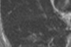
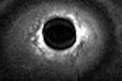
60-year-old woman with history of left mastectomy presents for follow-up MRI examination of right breast with normal mammogram and normal physical examination. Above, sagittal fat-suppressed image of right breast after contrast injection immediately before biopsy confirms presence of mass (arrow). Below, sagittal fat-suppressed image of right breast immediately after 9-gauge MRI-guided vacuum-assisted biopsy shows obturator (arrow) in high-signal hematoma obscuring biopsy site.
 |
The results showed that the histology based on MR biopsy was pure DCIS in 29 cases, and DCIS with possible microinvasion in five cases. Among the pure DCIS lesions, surgery revealed invasive carcinoma in 17% of the cases. This underestimation rate fell between the 11% reported at 11-gauge stereotactic vacuum-assisted breast biopsy and the 20% rate associated with 14-gauge automated core biopsy, the authors said.
Breaking down the results, the group found that 29 pure DCIS lesions were in 29 postmenopausal women with a median age of 56 years. The median lesion size was 1.5 cm and the majority (62%) was not masses. The median number of biopsy specimens obtained was 10. During surgery, invasive carcinoma was found in five cases.
 |
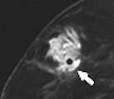
| Same patient as above. Histologic analysis of vacuum-assisted breast biopsy specimen shows ductal carcinoma in situ (DCIS) with solid architecture, intermediate nuclear grade, and central mucin (H and E, x40). Lee J, Kaplan JB, Murray MP, Mazur-Grbec M, Tadic T, Damir Stimac D, and Lieberman L, "Underestimation of DCIS at MRI-Guided Vacuum-Assisted Breast Biopsy" (AJR 2007; 189:468-474). |
In the five cases with possible microinvasion, the women had a median age of 46 with only one being premenopausal. The median lesion size was 2.5 cm and the MRI lesion type was nonmass in 80% of the cases. The median specimen number was 12. Surgical excision revealed invasive cancer in four cases.
The group offered several possible reasons for the underestimation rate. First, there is the high risk status of women undergoing MR breast biopsy, who are more likely to have invasive cancer. In addition, larger lesions (6 cm and higher) are associated with invasive cancer at surgery. Finally, MR biopsy historically has underestimated atypical ductal hyperplasia (ADH).
On the topic of larger lesions, "we suggest consideration of sentinel node biopsy during definitive surgery for women with larger lesions yielding DCIS at (MR biopsy)," they wrote. This would prevent a second operation after the biopsy in order to assess the axilla, they explained.
In an interesting footnote, Lee's group did not find that MR lesion type and kinetics acted as significant predictors of DCIS underestimation. Previous studies by Dr. Christiane Kuhl and colleagues at the University of Bonn in Germany have made a connection between biological markers and DCIS on MR.
By Shalmali PalAuntMinnie.com staff writer
August 16, 2007
Related Reading
Breast gamma imaging spots DCIS better than mammo, MR, August 13, 2007
MRI may aid diagnosis of ductal carcinoma in situ, August 10, 2007
MRI beats US for breast screening of at-risk women, but yields more biopsies, August 6, 2007
Mammography effectively detects recurrent DCIS postradiotherapy, July 27, 2007
Residual DCIS after treatment can be considered a pathologic complete response, July 13, 2007
MRI spots high-grade DCIS more often than mammography: study, June 4, 2007
Copyright © 2007 AuntMinnie.com