MRI has been unfairly labeled as insensitive for diagnosing ductal carcinoma in situ (DCIS), according to a breast MR expert. In fact, DCIS cases found on MR are reflective of an advanced biological disease profile and not necessarily overdiagnosis, said Dr. Christiane Kuhl.
Kuhl's group, from the University of Bonn in Germany, performed a prospective observational cohort study comparing MRI to mammography for DCIS diagnosis. In a talk at the 2006 RSNA meeting in Chicago, Kuhl explained that breast MRI has been pegged as having limited value because DCIS is a major cause of false-negative results.
In addition to comparing the modalities in this investigation, Kuhl's group asked if "MRI-detected DCIS differs from mammographically detected DCIS with regard to biological profile?" she said.
This study included more than 5,000 women who were referred to a tertiary care center for screening or diagnostic tests. They underwent bilateral mammography with at least two views and contrast-enhanced bilateral MRI. Only women with pure DCIS were included, so all cases of invasive or microinvasive carcinoma were excluded, Kuhl said. Of the 5,612 women, there were 137 women with pure DCIS, and the authors examined the modes of detection in these cases.
Among the 137 women, 50% of DCIS cases were found with mammography and MRI. Forty percent were only found with MRI versus 8% that were only mammographically positive. Both modalities failed to find DCIS in 2% of the cases.
Overall, breast MR was positive for DCIS in 90% of the cases compared to mammography, which was positive in 58%. Out of 11 DCIS cases that were found on mammography only, 78% were high-grade. However, of the 55 cases found only with MRI, 91% were high-grade disease, Kuhl said. MRI was false-negative in 7% of the cases and these were all nonhigh-grade DCIS.
In high-grade disease, breast MR's sensitivity was 98% compared to 49% sensitivity for mammography. In nonhigh-grade disease, MRI still had better sensitivity at 78% versus mammography's 71%. Finally, MR achieved 100% sensitivity in estrogen receptor-positive cases, Kuhl said, adding that more than half of the high-grade DCIS cases were not diagnosed by mammography.
"MRI offers a significantly higher sensitivity for diagnosing pure DCIS.... The additional 'only MRI-detected' DCIS exhibited histologic and immune-histochemical evidence of biologic aggressiveness.... We propose that additional MRI-detected DCIS do not constitute overdiagnosis," Kuhl and colleagues concluded in their abstract.
In response to an RSNA attendee's question, Kuhl acknowledged that the study did have some bias as this was not a population of women were who considered at high risk for breast cancer. Instead, these women underwent screening by both modalities by choice so could they could be better informed than the average population, she said.
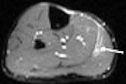
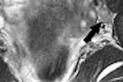
Last year, Kuhl authored an extensive paper on differential diagnosis in breast MRI for the journal, Magnetic Resonance Imaging Clinics of North America. She stated that "nonmass-like enhancement that follows the ductal system is the hallmark of DCIS in breast MR imaging" (August 2006, Vol. 14:3, pp. 305-328).
Other noteworthy aspects of DCIS on breast MRI that Kuhl recommended imagers keep an eye out for:
- About half of enhancing DCIS cases exhibit shallow enhancement; the other cases reveal early enhancement, but a washout time course is rare.
- DCIS diagnosis is based mainly on the assessment of lesion morphology, such as configuration and distribution.
- The enhancement pattern can be granular, stippled, or clumped.
- Most DCIS does not appear as a mass.
- Ductal or segmental enhancement on breast MR should be followed up with biopsy.
- If an intraductal cancer is large enough, internal enhancement can be evaluated with MRI.
Finally, Kuhl outlined other hallmarks of invasive cancer and how they related to breast MR protocol. "At kinetic analysis, the typical invasive cancer exhibits rapid and strong enhancement, followed by washout in the early postcontrast phase," she wrote. "In T1-weighted precontrast images and on nonfat-suppressed T2-weighted images, the typical invasive breast cancer exhibits low signal intensity equivalent to or lower than that of normal fibroglandular tissue."
By Shalmali Pal
AuntMinnie.com staff writer
January 12, 2007
Related Reading
Breast MRI measures biologic significance of DCIS, October 18, 2006
Radiation helpful in women with ductal carcinoma in situ, September 27, 2006
Breast MR falls short for microcalcifications, but CAD helps in malignancies, July 6, 2003
MRI best for familial breast cancer detection, December 16, 2005
Copyright © 2007 AuntMinnie.com