On prevertebroplasty MRI, the loss of signal intensity from the vertebral bone marrow space is indicative of bone marrow edema. This information is useful for determining which vertebrae should be treated. But according to a new study in Radiology, a direct connection between bone marrow edema and clinical outcome after percutaneous vertebroplasty (PVP) has not been established.
Dr. Noboru Tanigawa, Ph.D., and colleagues took a closer look at bone marrow edema patterns on pre-PVP MRI in 80 patients at the Kansai Medical University in Osaka, Japan. These patients were scheduled to undergo PVP for vertebral compression fractures resulting from osteoporosis.
In addition to MR, the patients underwent anterior and lateral x-ray of the thoracic and lumbar vertebrae and CT imaging. MR was done on a 1.5-tesla scanner (Signa, GE Healthcare, Chalfont St. Giles, U.K.) in 157 vertebrae (95 lumbar, 62 thoracic). The protocol included sagittal T1- and T2-weighted spin-echo sequences. A quadrature thoracolumbar spine coil was used.
The MR images were reviewed by Tanigawa's co-authors, Dr. Koshi Ikeda, Ph.D, and Dr. Naoto Omura, both neuroradiologists. Regions that exhibited signal intensity lower than that of fatty bone marrow on T1-weighted images and higher than fatty bone marrow on T2-weighted images were indicative of edema, according to the study's classification system.
Patients were grouped into the following categories:
- Group 1, type 1: bone marrow edema present in 50% or more of the vertebral body
- Group 2, type 2: bone marrow edema present in less than 50% of the vertebral body
- Group 3, type 3: no bone marrow edema
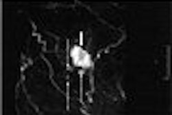
Tanigawa, or a fellow under his training, performed PVP with CT and fluoroscopic guidance. Pain level was evaluated with a visual analog scale (VAS) of 0 (no pain) to 10 (severe pain).
According to the results, there were 60 type 1 vertebrae, 28 type 2 vertebrae, and 69 type 3. Group 1 had 48 patients, group 2 had 14, and 22 were assigned to group 3. The authors reported a statistically significant difference between group 1 and group 3 in terms of the postprocedure VAS score (p = 0.037). The VAS score improvement (preprocedural score minus postprocedure score) was 4.6 in group 1, 3.7 in group 2, and 1.9 in group 3.
In group 1, 30 of 44 people experienced a pain reduction of at least 50%; eight of 14 had the same in group 2; and nine of 22 in group 3.
 |
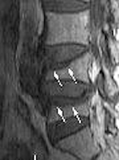
| Sagittal T1-weighted (above) and fat-suppressed T2-weighted (below) MR images of osteoporotic compression fracture of L4 and L5 vertebral bodies with considerable bone marrow edema pattern in a 74-year-old woman. Regions of low signal intensity (arrows) are present in the anterosuperior portion of L4 and L5 in image above and occupy less than 50% of the vertebrae. Regions of high signal intensity (arrows) are seen in the anterosuperior portion of L4 and L5 in image below. The high signal intensity regions occupied less than 50% of the vertebrae (group 2). |
 |
| Figures 2a, b. Tanigawa N, Komemushi A, Kariya S, et al. "Percutaneous Vertebroplasty: Relationship Between Vertebral Body Bone Marrow Edema Pattern on MR Images and Initial Clinical Response" (Radiology 2006; 239:195-200). |
"Our results suggest that the more extensive the bone marrow edema pattern is in the treated vertebrae, the greater pain relief provided by PVP will be," the authors stated. "All groups showed a statistically significant reduction in pain after the procedure, and pain reduction was significantly greater in group 1 than group 3" (Radiology, April 2006, Vol. 239:1, pp. 195-200).
Patients without bone marrow edema pattern are those with older, chronic compression, they explained. The use of PVP in this population remains controversial as PVP is credited for being most effective in acute compression factors.
Patients with greater bone marrow edema patterns on preoperative MRI would be more likely to benefit from PVP, the authors concluded. They acknowledged that their choice of MRI sequences was a limiting factor in this study. Future research should include short-tau inversion-recovery sequences as well as contrast enhancement.
By Shalmali Pal
AuntMinnie.com staff writer
May 18, 2006
Related Reading
New fractures occur sooner in adjacent vertebrae following vertebroplasty, January 24, 2006
Vertebroplasty packs radiation punch, but cement injector can soften blow, December 5, 2005
Copyright © 2006 AuntMinnie.com