SAN FRANCISCO - Damage to the brain stem after a traumatic head injury is likely to be fatal, according to an MRI study presented this week at the American Association of Neurological Surgeons (AANS) meeting.
Dr. Raimund Firsching presented the results of a seven-year study that included 200 patients who had sustained a traumatic head injury and had been in a coma for at least one day. Firsching and co-author Dr. Dieter Woischneck are from the Klinik fur Neurochirurgie at Otto-von-Guericke Universität in Magdeburg, Germany.
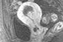
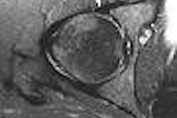
"We know that MRI can identify lesions when CT fails to do so," Firsching said. In previously published work, the authors analyzed the frequency and significance of traumatic brain stem lesions on MRI after severe head injury. They found that severe destruction of supratentorial white matter was not related to increased mortality, as long as the brain stem was spared. However, mortality was 100% when the lesion affected the pons or caudal portions of the medulla oblongata bilaterally (Neurological Research, March 2002, Vol. 24:2, pp. 145-146; Child's Nervous System, March 2003, Vol. 19:3, pp. 174-178).
For their current study, 200 patients underwent 1.5-tesla MRI, as well as CT, between 1996 and 2004. The location of lesions was correlated with mortality, duration of coma, and outcome.
The researchers found that 37% of the 200 patients had supratentorial lesions (grade I), while 20% had unilateral lesions of the brain stem (grade II). In addition, 22% had bilateral lesions of the mesencephalon (grade III) and 21% had bilateral lesions of the pons (grade IV).
They concluded that pontine and midbrain lesions seen on MRI, but not on CT, proved to be of "paramount prognostic relevance." In their study group, 92% of those with bilateral pontine lesions died. They also determined that the duration of the coma was linked to the severity of the head injury, particularly when the brain stem was involved.
"The locations of the lesions identified with MRI were highly and significantly correlated with mortality, duration of come, and outcome … at least three months after head injury (p ≤ 0.001)," the authors wrote in their AANS abstract. Firsching added that, in order to make the most of MRI in head trauma patients, neurosurgeons must work closely with neuroradiologists.
Commenting on the paper was Dr. M. Ross Bullock, Ph.D., from the Virginia Commonwealth University Medical Center in Richmond. He praised the researchers for their large study population, for performing MR studies within the first eight days of injury, and for demonstrating the connection between brain stem lesions and outcomes.
However, Bullock said that the study did have some weaknesses. Firsching and Woischneck did not attempt to differentiate between primary injuries and post\herniated changes, which can create brain stem lesions, he said. Bullock also said that the authors needed to look into the cost-benefit ratio of performing MR tests during a coma patient's first week in the intensive care unit.
Firsching acknowledged that in Europe, money is less of an issue than it is the U.S. "We are in a part of the world with heavily socialized medicine.... We have this bizarre situation where we can order elaborate neuroimaging without any cost to the patient or the department of neurosurgery," he said.
By Shalmali Pal
AuntMinnie.com staff writer
April 27, 2006
Related Reading
Doubling down: Raising the stakes of MRI patient safety, March 9, 2006
CT is the blanket modality for neuroimaging blunt injury patients, January 12, 2006
MRI finds long-term concussion effects, August 29, 2004
Copyright © 2006 AuntMinnie.com