An intra-arterial MR angiography (MRA) technique that has proved viable in animal studies has been successfully translated to humans, according to a new study. Meanwhile, another study has found that computer aided-detection (CAD) can help radiologists diagnose intracranial aneurysms on MRA more effectively.
In the first paper, Dr. Niels Zorger and co-investigators conducted a prospective study that evaluated the feasibility of intra-arterial MRA of the infrainguinal arteries for stenosis. Imaging was done before and after balloon angioplasty, and compared to digital subtraction angiography (DSA).
"We also aimed to show that a low dose of contrast agent could be used with this new technique," wrote the authors from the University of Regensburg in Regensburg, Germany (American Journal of Roentgenology, October 2005, Vol. 185:4, pp. 867-872).
Fifteen subjects with arteriosclerotic obstructive disease were included in this study. The study protocol began with an 11-cm 5-French sheath introduced antegrade of the ipsilateral common femoral artery. An initial DSA of the whole leg was done with half-strength contrast material.
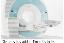
MR was performed on a 1.5-tesla unit (Magnetom Sonata, Siemens Medical Solutions, Erlangen, Germany) in addition to 3D MRA. A 30-mL solution of 5 mL of gadolinium diluted with 55 mL of a 0.9% saline solution was administered through the sheath. After MRA, balloon angioplasty was performed followed by DSA and another MRA exam.
The results showed that intraarterial MRA was technically successful in all 30 exams. Based on the grading system for peripheral MRA set by the American College of Radiology (ACR), the images were graded as excellent to good in 27 of the 30 intra-arterial MRAs.
On DSA, 64.4% of 180 segments were judged to be patent. For detecting significant stenosis in the femoropopliteal vessels, the sensitivity of MRA was 92.4% and the specificity was 91.7%. In the crural vessels, MRA's sensitivity was 91.9% and the specificity was 87.8%. In the complete leg, intra-arterial MRA achieved a mean sensitivity of 92.2% and a mean specificity of 88.6%.
Overall, "we confirmed the accuracy of intra-arterial MR angiography for detecting stenosis and treatment success after angioplasty," the authors stated. More importantly, they demonstrated that it was possible to use diluted contrast media, which not only protects patients from excessive dosage, but can also be more efficient.
"In our study, for one acquisition of a defined vascular region only, 2.5 mL of nonionic 0.5 mol/L of gadodiamide diluted with 27.5 mL of saline solution had to be injected intraarterially through the sheath," they explained. "Based on an 80-kg patient, theoretically 19 separate acquisitions of the treated, vascular region are possible ... enough for most interventional procedures."
MRA with CAD
In the second study, Dr. Toshinori Hirai and colleagues acknowledged that MRA is a widely accepted screening tool for intracranial aneurysms. But this exam produces a prodigious amount of data, the analysis of which can be time-consuming. In addition, it can be difficult for experienced readers to spot small aneurysms.
"Since the workload of radiologists will increase in the near future, the use of CAD may be necessary to increase true-positive findings and decrease false-positive findings," the authors wrote. "A CAD scheme has not been applied to the detection of intracranial aneurysms with (MRA)," they added (Radiology, November 2005, Vol. 237:2, pp. 605-610).
Hirai's group is from several Japanese institutions: Kumamoto University in Kumamoto, the University of Occupational and Environmental Health in Kitakyushu, and Kyushu University in Fukuoka. Another contributor is from the University of Chicago in Illinois.
For this retrospective review, MRA exams were culled for 60 patients with unruptured intracranial aneurysms and for 60 additional patients without intracranial aneurysms. MRI was done on a 1.5-tesla unit (Magnetom Vision, Siemens Medical Solutions) and 3D time-of-flight MR angiograms were obtained.
Of these 120 images, 28 from each group were selected for the observer performance study. The 15 observers consisted of neuroradiologists and general radiologists. Each read the MRAs on a monitor without CAD input and rated his or her confidence level as to the presence or absence of an aneurysm. The CAD output was superimposed on the MRAs and rated again. The CAD scheme was described in an earlier paper (Academic Radiology, October 2004, Vol. 11:10, pp. 1093-1104).
For all observers, the area under the ROC (Az) value was 0.931 without CAD compared to 0.983 with CAD. For the neuroradiologists, the Az value was 0.963 without CAD and 0.984 with CAD. For general radiologists, the Az value was 0.894 without CAD and 0.983 with CAD.
"The performance of general radiologists with CAD slightly exceeded that of neuroradiologists without CAD," the authors concluded. Among the patients with aneurysms, 10% benefited from CAD information. Eight percent of those without aneurysms benefited from CAD.
Tracking intracranial aneurysms with MRA can be tricky: It requires knowledge of intracranial anatomy, common locations for aneurysms, and the pitfalls associated with the modality, Hirai's group stated. The fact that both sets of patients saw benefits from CAD-enhanced MRA indicated an increase in observer confidence, whether they were general radiologists or brain imaging specialists, they added.
By Shalmali Pal
AuntMinnie.com staff writer
November 17, 2005
Related Reading
MRA shows good results in detecting coronary stenosis, March 7, 2005
3D contrast MRA may be optimal for evaluating aortic dissection, December 30, 2004
Whole-body MR permits rapid evaluation of lower peripheral arterial system, July 16, 2004
Copyright © 2005 AuntMinnie.com


