Animal models have been lacking in virtual colonoscopy studies, but not for lack of potential. Animal experiments can be useful for studying experimental contrast agents and imaging techniques, two important areas of VC research, in a highly controllable cohort. And the results can be verified by histology, rather than a reference standard designed to accommodate human needs.
At the University of Essen in Germany, Dr. Christoph Herborn, Dr. Fan Yang, Dr. Jörg Debatin, and colleagues performed MR colonography on a group of Wistar rats earlier this year. The study confirmed MRC's high sensitivity and specificity in data verified by histology. The group also solved a number of technical challenges to build a workable model for future efforts for colon imaging in animal models.
"Different modalities have been proposed and evaluated for (virtual colonoscopy)," Herborn said at the 2004 European Congress of Radiology. "Our experience with MR is that it has excellent soft-tissue contrast and very good temporal and spatial resolution." The contrast agents used with MRI generally have a better safety profile than those used with CT.
Finally, he said, improved results achieved with the latest gradient technologies appear to make MR the modality of choice, and its lack of radiation makes it ideal for screening.
"The aim of our study was to evaluate the dark-lumen contrast-enhanced MRC protocol for detection of colorectal polyps in a rat model," he said.
The researchers examined 14 male Wistar rats (age 12-15 months, mean 13.4 months) with MRC a year after the animals had undergone repeated enemas of N-methyl-N'-nitro-N-nitrosoguanidine, a toxic mixture that induces colonic adenomas over the course of several months.
"Twenty-four hours prior to MR exam, the animals were deprived of food -- only allowed to drink sweet water, which leads to colon cleaning," Herborn said. "The animals were anesthetized by injection of pentobarbital. In order to have spasmolysis, we injected them with (0.5 mg/kg) of scopolamine, which is Buscopan (Boehringer Ingelheim, Ingelheim, Germany). The animals then received a rather small saline enema at body warm temperature (37°C), and the protocol consisted of two sequences."
The radiologists acquired both plain and contrast-enhanced MR images on a 1.5-tesla MRI scanner (Magnetom Sonata, Siemens Medical Solutions, Erlangen, Germany) using a standard 30-cm knee coil. Supine images were acquired using two protocols: an unenhanced localizer scan composed of three image stacks in the coronal, sagittal, and transverse planes (TR/TE 500/20, FA 30° TA 30 sec.) and a contrast-enhanced T1-weighted 3D-VIBE (volumetric interpolated breath-hold examination) acquisition (TR/TE 4.5/1.5, FA 30°, 144 x 256 matrix, acquisition time 3:06 min., spatial resolution 0.6 x 0.5 x 1.2 mm). Contrast enhancement was achieved through intravenous administration of 0.3 mmol/kg Dotarem (Gd-DOTA, Guerbet, France), adjusted by body weight. To increase lesion conspicuity, the unenhanced MRI data was subtracted from the enhanced data.
 |
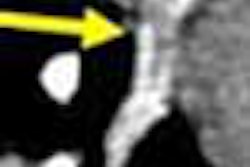
| (Left to right) The animal's chest and abdomen can be seen in the native unenhanced dataset, along with some motion artifact. Image quality improves slightly in early phase contrast, and significantly in late-phase contrast, four to five minutes after intravenous administration. Subtracting the native dataset from the early or arterial-phase dataset increases conspicuity of the colorectal lesion (arrow). All images courtesy of Dr. Christoph Herborn. |
Two radiologists examined multiplanar images, 3D reformatted images, and endoluminal views using Leonardo software (Siemens Medical Solutions), calling each lesion by consensus.
The group also assessed contrast uptake quantitatively in the colon wall and in the lesions via signal-to-noise (SNR) and contrast-to-noise (CNR) measurements, and compared the two using a Wilcoxon-Mann-Whitney U-test. The animals were sacrificed immediately after imaging, and the MRC results compared with histology.
 |
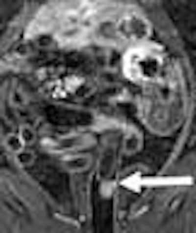
| Native dataset (left) again offers little detail, but strong contrast uptake in arterial-phase enhancement reveals a large lesion in the sigmoid colon. Data subtraction technique further increases lesion conspicuity. |
"Pathology was able to detect a total of 15 polyps in 9/14 animals, and five animals did not show any polyps." Herborn said. Of these, contrast-enhanced MRC detected 13/15 polyps measuring 6-13 mm (mean 7 ± 0.6 mm) in 8/9 rats, for a specificity of 87% and 100%, respectively. Two lesions (3 mm and 4 mm), missed in MRC but found in pathology, were not visible in a retrospective review.
 |
| Pathology shows colorectal lesion above the 6-cm marker on the ruler. |
Contrast-to-noise and signal-to-noise ratios increased significantly with the addition of contrast, from 69.4 ± 8.7 to 143.7 ± 11.3 (p < 0.03) for SNR, and from 41.2 ± 105.4 ± 14.1 (p < 0.01) for CNR. In the polyps, SNR increased from 78.2 ± 6.3 to 167.4 ± 17.7 (p < 0.02), while CNR increased from 45.4 ± 5.2 to 124.6 ± 11.2 with the addition of contrast. The uptake of contrast in the polyps was significantly higher than in the surrounding normal tissues (p < 0.04).
"We have a new and feasible rodent animal model for imaging adenomatous polyps," Herborn concluded. "The combination of dark lumen and bright colon wall in the protocol of contrast-enhanced MR colonography is a practical approach for detection of these lesions."
This improved enhancement appears to facilitate polyp detection, and needs further evaluation, "both in an experimental setting and with regard to diagnosis and treatment," he said. "The dark-lumen MRC exam must be further assessed in patients with respect to polyps."
The animal model appears to be well-suited for evaluating new gene-therapy techniques, he said.
By Eric Barnes
AuntMinnie.com staff writer
October 8, 2004
Related Reading
Digital subtraction enhances colonic lesions in MRC, July 7, 2004
New tagging, subtraction techniques aim for better compliance in VC, March 8, 2004
German group optimizes barium-sulfate tagging in MR colonography, January 22, 2003
Contrast agents herald new progress in MR lymphography, August 9, 2002
Copyright © 2004 AuntMinnie.com