The caffeine kick that jumpstarts the day for so many people could also boost the results of BOLD fMRI studies. Researchers from Northwestern University and Medical School in Chicago tested small doses of caffeine as a way to improve blood oxygen level-dependent (BOLD) signal response on functional MRI.
"Caffeine is a cerebral vasoconstrictor that can decrease the resting-state cerebral blood flow (CBF) in humans by as much as 20%-30%...we investigated the use of caffeine as an agent to decrease the resting CBF and increase the deoxyhemoglobin concentration in the baseline condition, allowing an overall increase in the BOLD response during the active condition," wrote Todd Parrish, Ph.D. in the journal NeuroImage (January 2002, Vol.15:1, pp. 27-44). Parrish’s co-authors were Todd Mulderink, a third-year medical student, and neurologists Dr. Darren Gitelman and Dr. M. Marsel Mesulam.
Eighteen healthy, right-handed Northwestern students were enrolled in the study, including 10 who were given caffeine, 4 who made up the control group, and 4 who were examined both with and without caffeine. All of the students were asked to refrain from ingesting any caffeinated food or drink 12 hours prior to imaging.
The MRI exams were done on a 1.5-tesla Magnetom Vision Plus (Siemens Medical Solutions, Iselin, NJ) equipped with echo-planar gradients. A Vac-Fix vacuum restraint pillow (Bionix, Malvern, PA) was used with the head coil. Superlab software (Cedrus, Phoenix) was used to process the behavioral responses. Both the functional and anatomic slices were oriented in an oblique axial plane.
Over a two-and-a-half-hour period, each group was studied in pre- and post-treatment phases using fMRI and a resting-state perfusion imaging method. The subjects were given either a 200-mg caffeine tablet (the equivalent of 2-3 cups of coffee) or a placebo.
For the functional exam, the single-shot echo-planar sequence acquired 6-mm slices with a 3-mm gap. Functional data analysis was done using Brain Voyager (Brain Innovation, Maastricht, the Netherlands) and included 3-D motion correction. The 3-D anatomic images were acquired in the same plane as the functional images.
 |
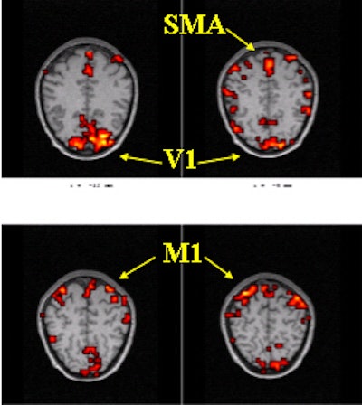
The areas of investigation were the left and right primary motor cortices (M1) and the primary visual cortex (V1). Above, a baseline fMRI scan during a visually cued motor task. Below, the same areas scanned after caffeine. Significant changes were seen in the signal change of the BOLD response. Images courtesy of Todd Parrish, Ph.D.
 |
The average BOLD signal amplitude response was determined from three areas of investigation, including the left and right primary motor cortex and the primary visual cortex. Additional analysis was done to measure the signal-to-noise ratio (SNR) in the nonactivated BOLD data, both before and after treatment.
After the administration of caffeine, the average percentage signal change of the BOLD response, relative to baseline, showed significant changes in both motor and visual areas, with the motor region exhibiting a 37% signal change. In addition, there was a notable decrease in the SNR following caffeine, with an average decrease of 4.4%. Finally, the average perfusion level decreased by 11%-13% without a change in performance as measured by reaction time, the group concluded.
"By exploiting the cerebral vasoconstrictive property of caffeine and its ability to reset the level of coupling between CBF and neuronal activity, it is possible to increase the BOLD signal response," they stated.
The study did have some limitations. First, the subject’s weight was not a factor in determining caffeine dosage. Also, whether or not caffeine was a part of the subject’s normal diet was not taken into consideration, although a regular coffee drinker might produce better imaging results, Parrish speculated in an e-mail to AuntMinnie.com.
Still, the group determined that the caffeine-enhanced signal increase could improve many aspects of BOLD fMRI studies, such as resolution, acquisition schemes, the robustness of activation, or more complex paradigms.
Parrish also suggested that this method could be of particular value in imaging cerebrovascular disease, a theory he said he is currently investigating.
By Shalmali PalAuntMinnie.com staff writer
May 14, 2002
Related Reading
Melatonin puts kids to sleep, makes MRI a dream, December 10, 2001
Java junkies exhibit different cerebral blood flow, November 28, 2001
Senna granules clear the way for better barium enemas, July 21, 2001
MR tracks effects of herbal treatment for prostate cancer, June 5, 2001
Blueberry juice enhances pediatric MR urography, April 26, 2001
Copyright © 2002 AuntMinnie.com