German researchers have found that F-18 sodium fluoride (F-18 NaF) PET/CT may provide "relevant information about the morphologic and functional properties of calcified plaque," according to a study published in the June issue of the Journal of Nuclear Medicine.
The study, from researchers based at the University Medical Center Hamburg-Eppendorf in Hamburg, also showed a strong correlation between radiotracer uptake and arterial wall calcification, with 88% of the lesions with F-18 NaF uptake showing calcification. "Unlike 18F-FDG or choline derivatives, 18F-sodium fluoride may provide new insights into the functional properties of calcified lesions," wrote lead author Thorsten Derlin, MD, and colleagues (JNM, June 2010, Vol. 51:6, pp. 862-865).
The retrospective study culled data from patients who received whole-body F-18 NaF PET/CT for the exclusion of bone metastases at the medical center between December 2008 and July 2009. Seventy-five patients with a mean age of 65.2 years were enrolled. Among the 48 women and 27 men, nine patients had a history of previous cardiovascular events.
Imaging protocol
PET and CT were performed with a PET/CT hybrid system (Gemini GXL 10, Philips Healthcare, Andover, MA), with F-18 NaF injected intravenously at a dose of 350 MBq ± 50. Patients first received whole-body nonenhanced low-dose CT of the whole body, followed by whole-body PET at 90 seconds per bed position at the head and thorax, and 60 seconds at the legs.
The analysis evaluated both lesions and arterial segments, with major arteries divided into the right and left common carotid arteries, ascending aorta, aortic arch, descending thoracic aorta, abdominal aorta, right and left iliac arteries, and right and left femoral arteries.
The researchers also classified arterial segments as CT-positive if at least one calcified lesion was detected by CT, and as CT-negative if no lesions were found by the modality. The same criteria applied to PET-positive and PET-negative results.
Arterial wall F-18 NaF uptake was seen at 254 sites in 57 (76%) of the 75 patients, with the greatest prevalence of uptake in the femoral arteries, followed by the abdominal aorta and thoracic aorta. Maximum standardized uptake values (SUVmax) ranged from 0.8 to 3.6.
The researchers also noted calcification at 1,930 sites in 63 (84%) of the 75 patients, with the highest levels of calcification in the abdominal aorta, followed by the femoral and iliac arteries.
F-18 NaF accumulation
"Prevalence of 18F-sodium fluoride accumulation was statistically significantly higher in lesions with extensive calcification than in those with only minor mineral deposition," the authors noted. "No statistically significant association between intensity of radiotracer accumulation (SUVmax) and calcification score was found. For 10 patients (13%), no arterial wall lesions were visualized by either PET or CT."
Of the 600 total arterial segments, 180 segments (30%) were PET-positive and CT-positive, while 160 segments (27%) were PET-negative and CT-positive. In addition, 10 segments (2%) were PET-positive and CT-negative, with 250 segments (42%) deemed PET-negative and CT-negative.
In the comparison of spatial correlation between vascular F-18 NaF uptake and calcification sites in the arterial segments, 31 (12%) of the 254 lesions with marked arterial wall F-18 NaF uptake did not show concordant calcification.
 |
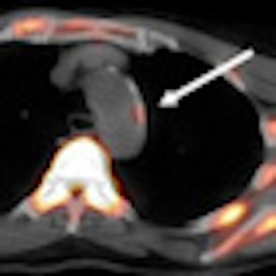
| Transaxial F-18 NaF images from a 76-year-old woman include CT (A), PET (B), and fused PET/CT (C) images of the aortic arch. F-18 NaF uptake in atherosclerotic lesion coincided with calcification, as indicated by the arrows. Images courtesy of the Journal of Nuclear Medicine. |
For the remaining 223 lesions with F-18 NaF uptake (88%), researchers found radiotracer accumulation and calcification. However, the study stated, "only these 223 (12%) of the 1,930 total calcification sites showed prominent 18F-sodium fluoride uptake in at least some part of the calcified plaque."
The authors cited several limitations of the study, including the lack of synchronization of PET image acquisition with both the cardiac cycle and the spontaneous respiration rate, which might have affected the visualization of arterial uptake, particularly with regard to the coronary arteries, the ascending aorta, and the aortic arch. Second, dynamic and delayed data could provide relevant additional information regarding the nature of arterial radiotracer uptake.
In addition, the researchers noted that limited spatial resolution of PET renders images subject to partial-volume effects, which might lead to an underestimation of tracer accumulation in atherosclerotic plaque, especially in smaller arteries.
By Wayne Forrest
AuntMinnie.com staff writer
June 11, 2010
Related Reading
CMS OKs F-18 NaF imaging coverage, March 8, 2010
F-18 fluoride PET/CT may better manage painful bone metastases, November 29, 2009
Sodium fluoride PET/CT changes management of foot pain patients, November 3, 2009
Copyright © 2010 AuntMinnie.com