A promising new molecular imaging technique may provide physicians and patients with a noninvasive way to obtain more information about endometrial carcinoma, according to a study published in the October issue of the Journal of Nuclear Medicine.
The study, led by Dr. Hidehiko Okazawa, professor in the division of medical imaging at Fukui University in Japan, used a specialized form of PET called estrogen receptor expression imaging for 22 patients with endometrial adenocarcinoma and nine patients with endometrial hyperplasia (a thickening of the uterine lining that is a risk factor for developing endometrial cancer) to evaluate diagnostic accuracy.
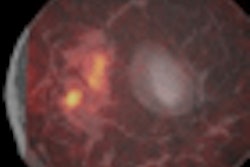
All patients underwent preoperative PET scans with fluorine-18 (F-18) fluoroestradiol, a tracer that has been successfully used in diagnosing breast cancer, and F-18 FDG, to compare differences in tracer accumulation.
The researchers confirmed that endometrial carcinoma reduces estrogen dependency with accelerated glucose metabolism as it progresses to a higher stage. By combining the two tracers, the group was able to use a new index of uptake ratio that can better predict pathologic stages and aggressiveness of tumors. The study results showed that the combined techniques had an accuracy rate of 86%.
Related Reading
UFE provides long-term relief of fibroid symptoms, June 30, 2009
Endometrial cancer radiation treatment increases second cancer risk, April 24, 2009
Survival odds better with RT for endometrial cancer, April 2, 2009
Lymphadenectomy and radiotherapy not useful for early endometrial cancer, December 15, 2008
Vaginal brachytherapy effective for endometrial cancer, June 3, 2008
Copyright © 2009 AuntMinnie.com