Indium-111-labeled octreotide imaging has demonstrated greater accuracy than CT or MRI scans in detecting somatostatin receptor-positive neuroendocrine tumors. However, anatomic localization of these tumors solely based on octreotide scans is problematic; thus, these procedures are usually performed in combination with CT or MRI.
A recent study from the Cleveland Clinic's division of radiology and department of molecular and functional imaging suggests that utilization of SPECT/CT in octreotide imaging can provide important diagnostic information to clinicians that is unavailable with planar whole-body scans.
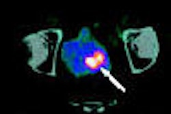
"Our standard imaging protocol includes full-body planar imaging six and 24 hours after tracer administration," said Dr. Lakshmi Ananthakrishnan, who presented the Ohio researchers' results at the 2006 RSNA conference in Chicago. "Physiologic uptake is seen within the thyroid gland, liver, gallbladder, spleen, and renal collecting system. Physiologic activity is seen within the GI tract as well, but usually only on the later images and sometimes is confounding to interpretation."
The scientists performed a retrospective study to determine the clinical utility of SPECT/CT localizing and characterizing octreotide-avid tumors. A review of 55 consecutive patients with suspected or pathology-proven neuroendocrine tumors who had undergone octreotide imaging was conducted by the team.
Each of the patients had a whole-body planar scan at six and 24 hours after octreotide injection. In addition, all patients had a SPECT/CT study performed on a Symbia T6 SPECT/CT system (Siemens Medical Solutions, Malvern, PA) 24 hours postinjection, according to Ananthakrishnan. The planar images were reviewed by the researchers first, then a subsequent review of the SPECT/CT and planar images was done.
"The clinical data was not known to the reviewer at the time of interpretation," Ananthakrishnan said. "The gold standard we used was histopathologic correlation on positive studies and clinical follow-up on negative studies," she noted.
For the 55-patient cohort, 31 patients presented no abnormalities on either the planar or SPECT/CT studies, and were asymptomatic on their follow up, Ananthakrishnan reported. One patient had negative findings on planar imaging and positive findings with SPECT/CT. The remaining patients in the study group, 23, had positive findings on planar imaging. From this subset, 18 had positive findings on SPECT/CT and five had negative findings.
"Most of these false positives seen on the planar images turned out to be GI activity, and one patient had activity in a hydronephrotic kidney," she said.
In the 18-patient subset that had positive studies on both whole-body planar imaging and SPECT/CT, Ananthakrishnan stated that the SPECT/CT studies provided incremental localization information. The researchers received histopathologic correlation on all but one patient, who had not had a tissue biopsy at the time of the presentation.
Ananthakrishnan reported that 16 of 17 patients were true-positive with eight neuroendocrine tumors, six carcinoids, one hepatocellular carcinoma, and one squamous cell carcinoma.
"We did have one patient that had a false positive with the thyroid gland on both planar and SPECT/CT imaging; a biopsy showed benign thyroid tissue," she said.
Ananthakrishnan said that SPECT/CT provided additional valuable information in 78% of the patients with positive studies, and that the modality reduced the number of false-positive results in 9% of their cases. In addition, SPECT/CT imaging visualized one lesion that was not observed on the whole-body planar images.
The group reported only a limited number of patients with a short follow-up time window, and said that no comparison had been made of SPECT to SPECT/CT. Based on their experience, however, they believe that SPECT/CT allows the localization of these kinds of tumors, which isn't possible with SPECT.
"We found that SPECT/CT is a valuable adjunct to planar imaging in localizing neuroendocrine tumors," Ananthakrishnan said.
By Jonathan S. Batchelor
AuntMinnie.com staff writer
January 12, 2007
Related Reading
SPECT/CT implementation: More than plug and play, June 7, 2006
Cardiac SPECT/CT fusion captures SNM's Image of the Year, June 6, 2006
SPECT/CT helps clarify ambiguous bone foci, February 9, 2006
A new use for SPECT/CT: Tracking stem cell migration, October 7, 2005
SPECT/CT pilots prostate brachytherapy in community setting, August 15, 2005
Copyright © 2007 AuntMinnie.com