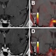
SAN FRANCISO - Serial SPECT imaging is an accurate and consistent way to measure cerebral hemodynamics after aneurysmal subarachnoid hemorrhage (SAH), according to a presentation this week at the Congress of Neurological Surgeons (CNS).
Arlid Egge, Ph.D., and colleagues from the University Hospital of Northern Norway in Tromsø imaged 32 patients who had been surgically clipped after SAH. They performed SPECT scans three to 26 days postoperatively (mean of 2.6 SPECT measurements). In addition, the patients underwent daily transcranial Doppler imaging. Finally, the Scandinavian Neurological Stroke Scale (SNSS) was used for daily clinical evaluation and scoring.
A single SPECT slice was divided into 16 symmetrical sectors, the researchers explained in their poster presentation. The automated template served to quantify regional cerebral blood flow. The group defined vasospasm using the following criteria:
Deterioration in clinical status that is based on SNSS and not attributable to other causes.
Transcranial Doppler measurements indicating a middle cerebral artery (MCA) to internal carotid artery (ICA) ratio of greater than four over three or more days.
On SPECT, corresponding, side-by-side sectors differing by more than 15% was defined as abnormal.
Summation of sectors on SPECT used to determine index for quantification.
"SPECT measurements in the acute phase after aneurysmal SAH follow a typical time sequence," the researchers concluded. In one case they described, a patient presented with a Fisher grade 2 bleed from a right-sided ICA aneurysm (grade II on Hunt-Hess scale). The SPECT measurement on day five was evaluated as normal. Measurements on days nine and 12 revealed increasing hypoperfusion in the right hemisphere.
In addition, the patient had an MCA/ICA ratio of seven and developed a left-side hemiparesis. On days 19 and 26, a hyperperfusion state occurred in the same region. After a year, a permanent defect developed and the patient remained severely disabled by hemiparesis.
"Hypoperfusion is followed by hyperperfusion, and this effect is most pronounced in patients with TCD and clinically evident vasospasm," the group concluded. "Repeated (SPECT) measurements are recommended."
Two years ago, the same group published results of a population-based study to assess the potential risk factors for SAH. The Tromsø health study surveyed nearly 30,000 people, and revealed that cigarette smoking, systolic blood pressure, and coffee consumption were independent risk factors for SAH (Journal of Neurology, Neurosurgery, and Psychiatry, August 2002, Vol. 73:2, pp. 185-187).
By Shalmali Pal
AuntMinnie.com staff writer
October 22, 2004
Related Reading
MR vs. CT? Stroke imaging hinges on more than modality, August 25, 2004
AAN issues transcranial doppler ultrasound guideline, May 12, 2004
ECG aberrations and hypomagnesemia linked in subarachnoid hemorrhage, May 18, 2004
Copyright © 2004 AuntMinnie.com


