The higher resolution of a prototype flat-panel volume CT scanner has enabled U.S. researchers to detect changes in the bone structure of girls with anorexia nervosa, even before declines in bone mineral density are evident.
Researchers from Massachusetts General Hospital (MGH) in Boston used flat-panel CT to assess trabecular bone microstructure of adolescent girls diagnosed with mild cases of anorexia whose bone mineral density (BMD) tests were normal. The researchers found that flat-panel volume CT revealed abnormalities of bone structure that were not identifiable with dual-energy x-ray absorptiometry (DEXA), quantitative ultrasound, and/or quantitative CT.
The study, published in the December issue of Radiology (2008, Vol. 24:3, pp. 938-946) and presented at the 2008 RSNA meeting in Chicago, determined that anorexia nervosa -- even in mild form -- in girls ages 12 to 18 can alter bone structure, placing these individuals at increased risk for fractures and osteoporosis as adults. Assessment of trabecular bone microarchitecture may improve the prediction of fracture risk and the capability to monitor responses to therapeutic intervention, suggested lead author Dr. Miriam Bredella, a musculoskeletal radiologist at MGH and assistant professor of radiology at Harvard Medical School in Boston.
The study was conducted between May and December 2007 and prospectively examined trabecular microarchitecture by using flat-panel volume CT. The researchers evaluated BMD by using DEXA in a cohort of 10 girls diagnosed with mild anorexia, compared with an age-matched control group of 10 girls with normal weight.
The girls with anorexia ranged in age from 13.8 to 18.2 years (mean, 15.9 years) and had 80% or more ideal body weight for their age. The control group ranged in age from 12.6 to 18.2 years (mean, 15.9 years) and had 90% or more ideal body weight.
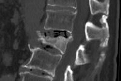
All subjects underwent anteroposterior radiography of the left hand to determine skeletal age. A CT of the nondominant distal radius was performed using a prototype flat-panel volume CT unit (Siemens Healthcare, Malvern, PA). The scanner has a bore diameter of 40 cm and uses an amorphous silicon detector material.
The hand of each subject was immobilized, with the wrist positioned using laser lights built into the scanner for alignment. Researchers obtained 580 projection images over a 360° angular span, conducted at 100 kV and 30 mA, with a pulsed x-ray source. The radiation dose at the distal radius was 0.027 mSv.
For each distal radius, a 3D oval region of interest was defined to cover a maximum area of bone without including any cortical bone. Calculations were made to determine the apparent trabecular bone volume fraction as a percentage, trabecular numbers, trabecular thickness, and trabecular separation.
Bone mineral density and body composition of the 20 girls were evaluated using DEXA (QDR 4500, Hologic, Bedford, MA), and parameters were obtained for BMD of the spine, hip, femoral neck, and total body; total bone mineral content; bone mineral content adjusted for height; fat mass; lean mass; and percentage of body fat. The researchers' analysis included correction for height, because DEXA tends to overestimate bone density in tall children and underestimate it in short children.
Linear regression analysis between trabecular structure parameters, BMD, and body composition was performed. The anorexic girls had lower body mass index, fat mass, and percentage of body fat compared to the control group, but there was no significant difference in lean mass. There also were no significant differences in BMD measurements of the spine and hip.
Trabecular structure measurements, however, were significantly different, the researchers discovered. Girls with anorexia had significantly lower values in apparent trabecular bone volume fraction, at 37%, compared with 46% for the control group. Their apparent trabecular thickness was 0.31 mm, compared with 0.39 mm for the control group, and the girls with anorexia had higher values of apparent trabecular separation, at 0.54 mm compared with 0.13 mm.
"Our data indicate significant deleterious effects on bone microarchitectural parameters in patients with [anorexia nervosa] even before changes in BMD are evident," Bredella said. "Reassuring values of BMD obtained by using DEXA may not reflect the true status of bone structure in this undernourished population and that alterations in bone structure may occur well before significant decreases in BMD become evident, especially when dealing with early or mild disease."
The research team pointed out that flat-panel volume CT allows the examination of a bone at high resolution (150-200 microns) with relatively low radiation exposure. The group also noted that trabecular structure parameters of the distal radius provide easy-to-measure markers that can be used to differentiate between adolescents with anorexia and normal-weight girls.
By Cynthia Keen
AuntMinnie.com staff writer
December 9, 2008
Related Reading
Menstruation key to bone rebuilding in anorexics, July 24, 2007
Skinny teens warned about osteoporosis risk, February 2, 2007
BMD recovery after anorexia based on weight gain and menstrual function, August 23, 2006
Copyright © 2008 AuntMinnie.com