SAN FRANCISCO - Volumetric CT scanning has evolved rapidly since the introduction of helical scanning and multiple detector rows. But even 64-detector-row CT can't acquire volumes thicker than about 8 cm in a single gantry rotation, which is not big enough to scan larger organs such as the heart without cobbling together data from multiple rotations. There are many ways to circumvent this limitation -- none of them optimal.
But a new research project aims to take CT imaging to the next level with a design known as inverse-geometry CT (IGCT), which incorporates multiple detector arrays for wide field-of-view imaging.
"Our long-term goal is to be able to image a thick volume, for example 15 cm or more, to make it scalable and to be able to image that entire volume in a single, fast rotation with uncompromised image quality," Dr. Norbert Pelc said Wednesday at the 9th International Symposium on Multidetector-Row CT, sponsored by Stanford University. "By that I mean no conebeam artifacts, isotropic, very high spatial resolution, and high dose efficiency."
Difficult applications such as cardiac CT angiography and perfusion studies would benefit enormously from the ability to image thicker volumes faster and with the highest resolution, Pelc said in his presentation.
 |
| All images courtesy of Dr. Norbert Pelc. |
There is not even a real prototype at this stage, only a tabletop device to demonstrate its scanning capabilities. But the project lacks neither talent nor support. Pelc and his co-investigators, including Dr. Samuel Mazin, Dr. N. Robert Bennett, Jongduk Baek, Arun Ganguly, Dr. Dominick Fleischmann and colleagues are from the departments of radiology, bioengineering, and electrical engineering at Stanford University in Stanford, CA. They are joined by Palo Alto-based detector array developer NovaRay; GE Healthcare of Chalfont St. Giles, U.K.; GE Global Research; and the National Institutes of Health in Bethesda, MD.
One way to acquire thick volumes in a single gantry rotation is with conebeam CT, which has a single, rotating point source and a 2D detector, Pelc said. However, flat-panel detectors have slower frame rates than conventional CT detectors, and the limitation of a single x-ray source and a circular scan path means there is not enough data for accurate reconstruction of the image data.
"These (conebeam) geometries have a fair amount of scatter that degrades the quality of data, and conebeam artifacts ... where you see the degradation in contrast and resolution at the upper and lower extents in this longitudinal reformation of the data," Pelc said. "And the severity of the artifacts increases as you increase your axial coverage on the system."
IGCT takes a different path. Instead of a single point source, it has large arrays of source points that can be rapidly turned on and off, combined with a smaller detector array. The in-plane field-of-view is determined by the size of this source array, while the field-of-view in a conventional CT system is determined by the detector array.
During the scan, "we very rapidly run through all of these point sources to collect data separately from each of them using a smaller detector array, and then rotate it about the patient," Pelc said. "Because both the source and the detector have the same axial extent, this system fundamentally has no conebeam artifacts. Because the detector array is smaller, we can use higher-technology detectors -- for example, photon counting systems -- and in addition (the design) also reduces the scatter content because at any instant in time we only have the small x-ray beam turned on."
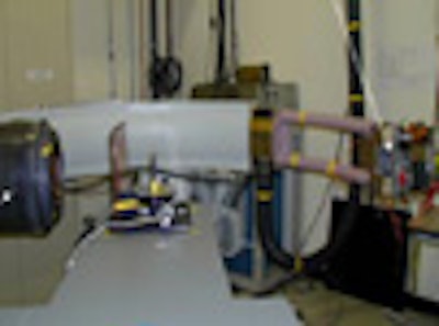
The detector technology comes from NovaRay, which developed an array of sources roughly 25 cm in diameter that can be turned on and off very quickly, combining it with a small detector array in a C-arm configuration. Originally designed for cardiac angiography, the Stanford researchers began developing the equipment with CT. They built a tabletop system that rotates the NovaRay C-arm laterally. For now the object to be scanned is placed on a turntable in front of the C-arm and rotated; there is no full-size prototype scanner yet, he said.
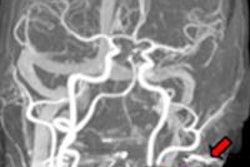
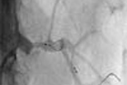
The initial results of the tabletop system in a phantom showed 5 cm of coverage with no conebeam artifacts. An image of a cadaveric inner ear specimen was imaged in a single rotation to show the higher spatial resolution compared to conventional CT. Truncation artifacts could be seen in a porcine heart imaged inside a phantom -- the 5-cm coverage of the tabletop system was insufficient for the size of the specimen.
 |
| IGCT eliminates conebeam artifacts, as shown in tabletop IGCT imaging demonstration above. Below, cadaveric inner ear scan on tabletop IGCT was compared to conventional MDCT scan. |
 |
A larger field-of-view can be obtained by at least two ways, Pelc said. One approach is to use three detector arrays, with each detector acquiring a different portion of the fanbeam. The data are combined to synthesize an effectively larger fanbeam and cover the full field-of-view.
The other approach for getting a large field-of-view is to build a large source array -- and it's technically difficult to build a source 100 cm long with a scanned electron beam, he said. "In collaboration with GE, we're investigating the approach of putting multiple pulsed sources in a single vacuum chamber," he said. "Opposite it would be a smaller 2D array of high-speed detectors, and the system will be mounted on a gantry."
 |
| In multisource IGCT, each portion of the fanbeam is smaller, but combined the fanbeam is larger than in conventional MDCT. The image below simulates the acquisition of 10 cm of coverage using a conventional conebeam scanner; underneath it is the simulated IGCT scan. With three sources in the z direction the conebeam artifacts are eliminated. Coverage can be scaled to an arbitrary thickness by choosing an appropriate number of sources. |
 |
The system concept also features a "virtual bow tie" filter that preshapes the intensity distribution of the fanbeam before it gets to the patient, modulating the intensity of the x-ray beam up and down to take advantage of the noncircular shape of the human body, Pelc said.
"With the multisource system you have an additional freedom in that as you pulse the sources you are free to have different intensities across that distribution," he said." And since each source is illuminating only a small portion of the object, you can effectively synthesize an optimal bow tie that changes the intensity distribution across the fanbeam as you rotate to take into account the nonsymmetric shape of the patient."
The goal of the project is the ability to scan an entire organ with fast rotation, and a simple point source rotation about the object will not suffice. For this a new approach to CT design is needed, one that goes beyond the use of a single point source rotating around an object. The group's solution to these challenges is to use a larger array of point sources combined with a smaller detector array.
"Our results are promising," Pelc said. "It's a technology that can be scaled to an arbitrary slab thickness, does not have conebeam artifacts, can deliver isotropic and homogeneous spatial resolution across a higher volume, and offers excellent dose efficiency, partly as a result of the virtual bow tie."
A detailed explanation of IGCT has just been published in Medical Physics (June 2007, Vol. 34:6, pp. 2133-2142), Pelc said.
By Eric Barnes
AuntMinnie.com staff writer
June 14, 2007
Related Reading
256-slice CT brings new possibilities in head imaging, May 28, 2007
Image postprocessing boosts performance of multidetector CT coronary angiography, May 31, 2007
Dual-source coronary CTA images the calcium-burdened, April 13, 2007
Copyright © 2007 AuntMinnie.com