VIENNA - The sound bites have barely stopped resonating from the sound and the fury of salvos fired off this week by proponents and detractors of CT as a lung cancer screening tool. Meanwhile, a study unveiled by researchers from Spain at this week's European Congress of Radiology (ECR) suggests that the current imbroglio swirling around CT screening may be only Sturm und Drang, at least when compared to the use of integrated PET/CT for lung cancer diagnosis.
According to the scientists from the department of radiology at the University Hospital La Paz in Madrid, Spain, PET/CT has several advantages over PET and CT for diagnosing lung cancer:
- The simultaneous acquisition of PET and CT studies provides a precise correlation between the anatomic and molecular data, which increases diagnostic accuracy.
- The use of CT to correct attenuation has allowed an improvement in PET image quality and reduced its data-acquisition time.
- Standardized uptake value (SUV) measures are more easily reproduced in PET/CT systems than in PET devices.
Over a 26-month period, the group examined 79 patients (69 men and 10 women; mean age of 62 years), who were referred to the study on the basis of imaging findings on a chest radiograph.
"The imaging findings on chest radiography considered suspicious of lung cancer were: pulmonary mass, atelectasis, mediastinal mass, and pneumonia without response to antibiotic treatment," the authors wrote in their electronic poster presentation.
Patients who had renal, hepatic, or other oncologic disease were excluded from participating in the study, as were patients presenting with an HIV infection, positive graving test, iodinated contrast allergy, or a previous diagnostic CT for lung cancer staging.
All patients in the study had an unenhanced low-dose CT for PET attenuation correction, an FDG-PET study, and a contrast-enhanced full-dose CT performed on a four-slice Discovery LS PET/CT (GE Healthcare of Chalfont St. Giles, U.K.).
According to the researchers, all patients fasted for six hours prior to the PET/CT exam, and their blood sugar levels were checked to ensure that there was no hyperglycemia. They received a standard 370 MBq intravenous dose of FDG approximately one hour prior to scanning, which was conducted from the base of the skull to the midthigh at five minutes per table position in the transverse field-of-view and caudocranial acquisition direction.
The diagnostic-level, contrast-enhanced CT was acquired following a 140 mL intravenous injection of iobitridol (Xenetix 300, Guerbet, Roissy, France) via an automated injector (XD 5500, Ulrich Medical Systems, Ulm, Germany) at 3 mL/sec. The data from this exam was resized from a 512 x 512 matrix to a 128 x 128 matrix to match the PET data and fuse the scans.
"The PET datasets were reconstructed iteratively with an ordered subsets expectation maximization algorithm (OSEM, segmented attenuation correction, and with the CT data," the authors wrote.
A nuclear medicine physician and a radiologist, both of whom had more than three years of experience with PET/CT, evaluated and interpreted the images in consensus.
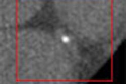
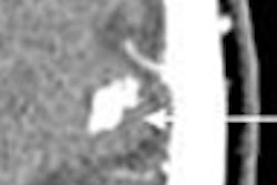
The hybrid modality correctly classified 71 of the patient cohort as having neoplastic disease: 27 had adenocarcinoma, 25 had non-small cell lung cancer (NSCLC), 14 had squamous cell carcinoma, and five patients had small-cell lung cancer. The other eight patients were false-positive because of inflammatory or infectious processes that mimicked lung cancer (three abscesses, three tuberculosis, and two pneumonias), the authors reported.
For 10% of the patient cohort with neoplastic disease, the PET/CT exam staged their lung cancer at IIIA or lower, which allowed them to undergo surgical resection or radiotherapy treatment. The remaining 90% of the patients with lung cancer were staged at IIIB or higher and received chemotherapy or symptomatic treatment.
"In our experience, integrated PET/CT may decrease radiation dose, diagnosis and staging delay, and improve cost-effectiveness compared with CT with intravenous contrast agents and PET performed independently; and could avoid other diagnostic procedures," the scientists wrote.
By Jonathan S. Batchelor
AuntMinnie.com staff writer
March 10, 2007
Related Reading
Italung-CT results show efficacy of lung cancer screening, March 9, 2007
CT screening may not improve lung cancer survival, March 7, 2007
Overdiagnosis may plague CT lung screening results, especially in women, November 29, 2006
PET, CT CAD scheme differentiates benign from malignant lung nodules, November 28, 2006
PET, PET/CT help determine RFA outcomes in lung cancer treatment, May 12, 2006
Copyright © 2007 AuntMinnie.com