Owing to its superior characterization of soft tissues, MRI is the modality of choice in head and neck imaging, and is likely to remain so. But the more CT technology improves, and the busier MR scanners get for imaging tasks not easily duplicated, the more CT is being used for head and neck applications.
"The closer you are to the skull base, the more MRI is preferred," said Dr. Nancy Fischbein, associate professor of radiology at Stanford University in California.
In California, with its large population of nasopharyngeal cancer patients, "MR is king because you're not going to see things like subtle clival marrow infiltration on a CT scan no matter how good your technique is," she said. On the other hand, "the more likely you are to have issues with motion degradation, the more CT is preferred."
CT is particularly well-suited to imaging vocal cord pathologies, laryngeal cancers, and airway compromise, Fischbein said at the 2005 International Symposium on Multidetector-Row CT in San Francisco.
And when weighing the use of a particular modality, it's important to consider the standard caveats that apply to the question of MR versus CT, such as contrast allergies, claustrophobia, and the like. The decision may ultimately come down to the "art of radiology," she said.
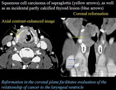
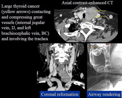
Axial images are the workhorses of MDCT head and neck imaging, and are often sufficient to make the diagnosis, Fischbein said. But coronal and sagittal reformations can be very helpful for some locations, notably the floor of the mouth, the larynx, the trachea, and the thyroid. Even treating models may have a role, particularly in airway imaging, she said.
"Another rule of thumb is that it's best to simply get in the habit of doing sagittal and coronal reformations for all cases," Fischbein said. "Otherwise you won't have them when you want them, and really it only takes the techs a moment once they get used to it."
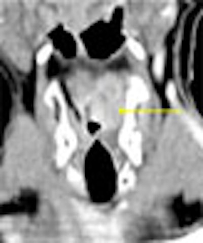
Projecting CT images of a patient with a right tonsillar abscess, Fischbein noted that while the axial image clearly depicts a peripherally enhancing necrotic puss collection, the coronal view adds potentially useful information.
 |
The coronal reformation can help surgeons "really understand the superior and inferior extension, the relationship to the nasopharynx, to the soft palate," she said, adding information that can aid in "planning their surgical drainage and make sure they don't leave puss pockets behind."
Another patient had a squamous cell carcinoma on the floor of the mouth, and lymph node metastases. Though the primary tumor was visible on the axial images, the sagittal reformations offered a better view of the mass at the floor of the mouth, while the coronal reconstructions provided a complete view of the nodal anatomy of the neck, Fischbein said.
 |
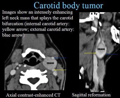
"These images can help us to detect additional nodes we might not have been sure of," she said. "Sometimes we might wonder if we're looking at focal necrosis or a fatty hilum. Of course you can … measure Hounsfield units, but sometimes just looking at a node in multiple planes tells you whether you're looking at focal necrosis or a fatty hilum."
 |
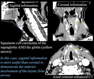
With regard to the larynx and airway, assessing the patient's ability to perform dynamic maneuvers can help the laryngologist determine the degree of laryngeal dysfunction, the prognosis for voice rehabilitation, and more.
 |
"With MR in a perfectly cooperative patient one gets a beautiful look at the false cords, the periglottic fat, the true vocal cords, and the laryngeal ventricle, and there are some people that have done dynamic laryngeal imaging with MR," Fischbein said. On the other hand, with MR scanners in high demand, phonation studies can be very time-consuming "when you've got acute strokes and other patients that you'd like to get into the MR scanner," she said.
MDCT is a powerful technique that can be used instead of MRI to address many questions related to the soft tissues of the head and neck, "in particular vocal cord pathology, laryngeal cancer and airway compromise," Fischbein concluded.
By Eric Barnes
AuntMinnie.com staff writer
November 18, 2005
All images courtesy of Dr. Nancy Fischbein.
Related Reading
Multiple modalities show similar results for initial staging of head and neck cancer, June 23, 2005
MDCT protocols weighed for cancers of larynx, hypopharynx, November 3, 2004
PET/CT shows restaging power for head and neck tumors, March 30, 2004
Scintigraphic sentinal node mapping cost-effective for early head and neck cancer, October 31, 2003
Chemoradiation study aims for 80% survival in laryngeal cancer, February 17, 2003
Copyright © 2005 AuntMinnie.com

