Images from a prototype 256-slice conebeam CT scanner offered complete volumetric data from a beating heart in a single gantry rotation, and without cardiac motion artifacts, researchers from Japan reported.
In a study using small pigs, the group from Chiba University Graduate School of Medicine and the National Institute of Radiological Sciences in Chiba, Japan, demonstrated segmented left ventricle (LV) perfusion combined with selective coronary arterial injection, displaying the LV enhancement three dimensionally.
"Information of the segmented left ventricular area supplied by every coronary artery is potentially useful for strategy and practice of percutaneous coronary intervention, coronary artery bypass surgery, or percutaneous transluminal septal myocardial ablation (PTSMA)," wrote Dr. Nobusada Funabashi and colleagues in a study published in Heart (October 2005, Vol. 91:10, pp. 1349-1351).
Myocardial contrast echocardiography with intracoronary contrast injection can be used to evaluate the segmented left ventricular area supplied by a coronary artery, but the procedure is invasive, and the ability to evaluate myocardial enhancement by contrast ultrasound may depend on the skill of the investigator, the researchers wrote.
For the study, two pigs weighing 20 kg each were anesthetized with isoflurane, and catheters were positioned in the left anterior descending (LAD) branch of the coronary artery in pig 1 and the left circumflex branch (LCx) in pig 2, via the femoral arteries. Heart rates for both animals ranged from 70-80 beats per minute, and 10 mL of iodinated contrast material was injected (300 mgI/mL). Scanning continued for 25 seconds for the experiment.
Images were acquired on a prototype 256-detector conebeam CT scanner (Athena, Sony-Toshiba) at 120 kVp, 200 mAs, 256 x 0.5-mm collimation, and 1-sec gantry rotation and exposure time.
Sixteen-slice CT images were also acquired for comparison on the group's regular Somatom Sensation 16 scanner (Siemens Medical Solutions, Erlangen, Germany) at 120 kVp, 250 mAs, 16 x 0.75-mm slice collimation, and 0.42-sec exposure and gantry rotation time, according to the researchers.
The radiation dose was 100 mSv for 25 seconds. Images were reconstructed at 468 x 0.468 x 0.500 mm3 with a 0.5-mm reconstruction increment along the z axis and a matrix size of 512 x 512 x 256, using standard body kernels (FC10), the authors wrote.
After two seconds, only the coronary arterial trees of LAD (pig 1) or LCx (pig 2) could be seen without myocardial enhancement, they found. At five seconds, segmented myocardial enhancement of LAD and LCx could be observed with the coronary arterial trees.
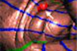
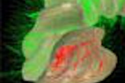
"We selected the most static images of the LV at around five seconds after contrast injection, so as to avoid any gaps from cardiac motion artifact," they wrote. "Axial source images clearly revealed segmented LV myocardial enhancement of the anterior and apical wall and interventricular septum (IVS) in pig 1, and the lateral and posterior wall in pig 2. Volume-rendered images from the anterior and the left anterior views showed only the anterior and apical wall and IVS portion of the LV myocardium supplied exclusively by the LAD in pig 1 and the lateral and posterior wall of LV myocardium supplied by the LCx in pig 2, together with the coronary artery; the segmented LV area could be easily recognized three dimensionally."
 |
 |
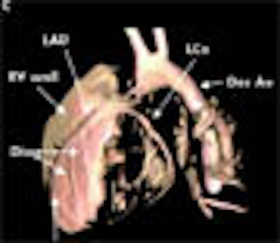
| Volume-rendered reconstruction images of porcine hearts using enhanced 256-slice high-speed conebeam CT acquired five seconds after injection of contrast material from the anterior view (A, B, above) and the left anterior view (C, D, below) revealed only the anterior and apical wall and interventricular septum (IVS) portion of the left ventricle (LV) myocardium, anterior wall of right ventricular myocardium supplied exclusively by the left anterior descending coronary artery (LAD) in pig 1 with LAD injection (A, C), and lateral and posterior portion of LV myocardium by the left circumflex artery (LCx) in pig 2 with LCx injection (B, D). The right coronary artery was visualized from the backflow from the catheter located in the LAD in pig 1(A) as well as the aorta. Asc Ao, ascending aorta; Des Ao, descending aorta; Diag; diagonal branch. Images from Heart 2005;91:1349-1351; doi:10.1136/hrt.2004.045997, used with permission of BMJ Publishing Group Ltd and British Cardiac Society © 2005. |
 |
 |
The prototype CT technique provides the advantage of whole-heart imaging in a single gantry rotation without gated acquisition, the authors wrote.
"Two plans can be envisaged for the future," they wrote. "Firstly, the combination of a selective angiography system with the CT system in which the CT acquisition with selective coronary artery injection can be achieved without any movement of the patient. In fact, this angiography system with multislice CT is already in use. Using the new system in which an angiography system is combined with a new 256-slice CT, it is possible to obtain the same segmented LV enhancement."
The second potential application is noninvasive 256-slice CT imaging combined with intravenous contrast injection. Even though this method does not allow for segmentation of the LV myocardial enhancement, 3D perfusion of the entire myocardium can be evaluated noninasvely, which is not possible with 16-slice CT scanners.
"Using this technique, continuous 25-second acquisitions can be performed yielding quantitative myocardial perfusion information by using time-density curves of myocardium on three-dimensional images," the authors wrote.
More research is needed to overcome potential problems, such as a high radiation dose and use of the catheter without complications, they noted. But the technique could potentially lead to new applications and diagnoses not only in the heart, but in the liver, kidney, and vasculature.
By Eric Barnes
AuntMinnie.com staff writer
October 24, 2005
Related Reading
CT nips at MRI's heels in LV function study, June 29, 2005
MDCT maybe equivalent to MRI, tops echo and SPECT for heart function, February 15, 2005
Contrast delivery key in coronary CTA, September 7, 2005
Multislice CT reveals previous plaque rupture in coronaries, June 14, 2005
CTA seen as accurate complement to invasive angiography, May 24, 2005
Copyright © 2005 AuntMinnie.com