Dialing down the x-ray tube current and activating tube-current modulation made a huge difference in the radiation dose for children undergoing coronary CT angiography (CTA), according to a new study in the American Journal of Roentgenology. The reduced-dose acquisition protocols had no significant negative effect on image quality and were, in fact, hardly noticeable, researchers said.
The retrospective study shows how two simple steps can provide safer heart scans for children, the patients most sensitive to being harmed by exposure to ionizing radiation.
"Only 43% of institutions adjust their CT techniques when examining children," wrote Dr. Christopher Herzog, Dr. U. Joseph Schoepf, and colleagues from Johann Wolfgang Goethe University in Frankfurt, Germany, and the Medical University of South Carolina in Charleston. "Several authors have recommended reducing the tube current-time product or the tube potential or both as a function of patient size, with the goal of obtaining constant diagnostic quality and image noise at reduced radiation. However, selecting a tube current that will yield acceptable image quality with the lowest possible radiation is still challenging," they said (American Journal of Roentgenology, May 2008, Vol. 190:5, pp. 1232-1240).
The study assessed the effect of weight-based scanning protocols and automatic tube-current modulation in children with congenital thoracic cardiovascular abnormalities, comparing the results of cardiac ultrasound, angiography, or surgery. The researchers examined data retrospectively from 68 consecutive pediatric patients (41 male, 27 female) who were referred for known or suspected congenital cardiovascular anomalies of the thorax.
All exams were performed after IV injection of 2 mL/kg of body weight of 300 mg I/mL of iohexol (Omnipaque 300, GE Healthcare, Chalfont St. Giles, U.K.) diluted 2:1 with 0.9% saline. Rather than breath-hold imaging, 37 of the youngest patients were scanned using the free-breathing technique to avoid the need for intubation or sedation.
Of the 68 patients, 38 were imaged using a 64-slice multidetector-row CT (MDCT) scanner (Somatom Sensation 64 Cardiac, Siemens Healthcare, Malvern, PA) at 64 x 0.6-mm collimation, z-flying focal spot technique, 0.33-second rotation, and pitch of 1.5, the researchers wrote.
For the 64-slice images, the tube voltage was adjusted to each patient's weight: 80 kVp for those weighing 15 kg (33 lb) or less (n = 17). Patients weighing more than 15 kg were scanned at either 100 kVp (n = 9) or 120 kVp (n = 12) at the discretion of the physician prescribing the scanning parameters. The patients were scanned patients using commercially available tube-current modulation software (Siemens CareDose 4D).
The other 30 patients underwent 16-slice MDCT (LightSpeed, GE) acquired at 16 x 0.625-mm collimation, 0.5-second rotation time, pitch of 1.5, 120-kVp tube voltage, and a weight-adapted tube current ranging between 32 and 110 mAs. The researchers calculated mAs, volume CT dose index (CTDIvol), and dose-length product (DLP) values, calculating the effective dose from these measures.
Image reconstruction used an individually adapted field-of-view of 512 x 512, and images were reconstructed at 0.75- and 3-mm-thick sections with increments of 0.4 mm and 3 mm, respectively.
Two experienced readers blinded to the scanning parameters and patient characteristics examined the data on a 3D-enabled workstation (Leonardo, Siemens) with a standardized window level of 100 H and window width of 700 H. They rated diagnostic image quality on a five-point scale, with 5 representing excellent diagnostic quality.
On the scale, the results showed mean diagnostic quality for 64-slice MDCT of 3.6 ± 0.4, with mean image noise of 8.9 ± 4.5 HU. Results with 16-slice MDCT were not significantly different with regard to diagnostic quality (3.6 ± 0.4; p = 0.97) or image noise (9.1 ± 2.8 HU; p = 0.31).
 |
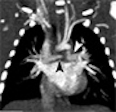
| Images show three different examples of congenital vascular abnormalities of chest evaluated with 64-slice CT with use of automatic tube current modulation. Oblique coronal maximum intensity projection images (upper row, above) and transverse section images (lower row, above) of patients scanned at 80 kVp and 100 kVp. Image set A (above) shows stenotic pulmonary artery (black arrowheads) in 6-month-old girl. Note difference in vessel caliber between right and left pulmonary arteries (white arrowhead). Image set B (below) shows left lower pulmonary vein (llPv) draining (black arrows) in right (rA) instead of left (lA) atrium in 2-year-old boy. ulPv = upper left pulmonary vein, rV = right ventricle, and lV = left ventricle. Image set C (100 mAs, bottom) shows tetralogy of Fallot with large septal defect (white arrows), overriding ascending aorta (aA), and stenotic pulmonary artery (Pa) in 6-month-old boy. |
 |
 |
| "Pediatric Cardiovascular CT Angiography: Radiation Dose Reduction Using Automatic Anatomic Tube Current Modulation," Christopher Herzog, Denise M. Mulvihill, Shaun A. Nguyen, Giancarlo Savino, Bernhard Schmidt, Philip Costello, Thomas J. Vogl and U. Joseph Schoepf (AJR 2008; 190: 1232-1240). |
In addition, scanning with automatic tube-current modulation significantly (p < 0.05) reduced the tube current time-product compared to scanning without automatic tube-current modulation (-57.8%/54.1/128 mAs) or with 16-slice CT (-47.9%/54.1/104.37 mAs), respectively.
While the mAs values were significantly (p < 0.05) lower at 80 kVp compared to 100 kVp or 120 kVp scans, image quality and image noise were not significantly different (p = 0.24). Agreement between MDCT and clinical findings was excellent. Moreover, all cardiovascular defects that had been documented on MDCT scans were correlated with cardiac sonography, the authors noted.
 |
| Comparison of dose and mAs values from "Pediatric Cardiovascular CT Angiography: Radiation Dose Reduction Using Automatic Anatomic Tube Current Modulation," Christopher Herzog, Denise M. Mulvihill, Shaun A. Nguyen, Giancarlo Savino, Bernhard Schmidt, Philip Costello, Thomas J. Vogl and U. Joseph Schoepf (AJR 2008; 190: 1232-1240). |
Agreement with surgery was 100% (95% confidence index [CI], 86.7%-100%), and also 100% (95% CI, 79.4%-100%) for catheter angiography. Good interobserver agreement was also seen for quality scoring (k = 0.76) and for detecting cardiovascular defects (k = 0.73).
"Evaluation of a sizable cohort of consecutive pediatric patients undergoing cardiovascular 64-(slice) MDCT shows that substantial reductions in radiation exposure can be realized by automated tube-current modulation techniques without sacrificing diagnostic quality," Herzog and colleagues wrote. "Use of 120 kVp for pediatric cardiovascular 64-(slice) MDCT incurs relatively higher radiation exposure, but does not significantly improve diagnostic quality compared with CT acquisition with lower tube potential. Thus, lower tube voltage settings appear recommendable for this patient population."
In line with previous studies, the researchers found that diagnostic quality is not impaired and image noise is only slightly increased if automated tube-current modulation is used.
However, the higher mean dose values seen in "virtual" 64-slice CT scans (i.e., those simulated to have been performed without automatic tube-current modulation) compared to actual 16-slice CT values reveals a slight overestimation of dose savings when 64-slice CT values are compared to default reference values, they wrote.
Noise does increase when the x-ray tube potential is reduced, they noted. However, contrast-to-noise ratio is the primary determinant of CT image quality, so noise is less important if the level of contrast is high enough and increases in line with tube-current reductions.
"In the present study, significant differences in the effective radiation dose were observed for 120 kVp compared with 100- and 80-kVp scans, respectively, whereas image noise and quality scores were comparable," Herzog and colleagues wrote. "In pediatric patients weighing ≥ 15 kg, beam energy of 100 kVp thus appears preferable over 120 kVp."
They cautioned that reference dose values for different kVp levels were derived exclusively from simulated CT exams, with limited value as a reference standard. Ethical considerations would prohibit any real measurements, the group wrote.
Other limitations of the study were seen in its small cohort and retrospective design, which did not allow for the creation of weight-dependent cutoff values for different beam energies, the group noted. Radiation doses were not directly measured but calculated based on the dose-length product, which has previously been shown to be accurate. Finally, scanning parameters such as collimator thickness, pitch, and gantry cycle time on radiation dose were not considered, and patients too young for the breath-hold technique were not assessed separately.
"In pediatric cardiovascular CT of the chest, automated tube-current modulation combined with low tube voltage leads to significantly decreased radiation dose while image quality is maintained," the authors concluded. "Standard tube potentials as they are used in adults tend to increase radiation in children without significantly improving image quality."
By Eric Barnes
AuntMinnie.com staff writer
May 21, 2008
Related Reading
Fetal echocardiography detects major but may miss minor heart defects, March 28, 2008
Dose studies delve into coronary CTA, March 8, 2008
Pediatric CT dose drops with breast shields plus tube modulation, February 26, 2008
Coronary CTA study aims for lowest dose, January 11, 2008
Increased use of CT scans raises risk of higher radiation exposures, November 29, 2007
Copyright © 2008 AuntMinnie.com