The upcoming introduction of a 256-slice CT scanner promises to make major contributions to CT instrumentation, according to a Japanese radiologist who is working with a prototype of the system. From 4D cardiac imaging to CT digital subtraction angiography, 256-slice CT may result in a host of new clinical applications.
Dr. Kazuhiro Katada, chairman of the department of radiology at Fujita Health University School of Medicine in Japan, discussed his experiences with 256-slice CT at Stanford University's International Symposium on Multidetector-Row CT in June. He also discussed the technology in an interview with AuntMinnie.com during the San Francisco meeting.
Katada has been involved in CT development since 1975, first with Hitachi Medical and since 1986 with Toshiba Medical Systems. He has worked on more than 20 prototype machines, and in many cases has been the first clinician to work with new Toshiba prototypes, such as the company's 64- and 256-slice systems.
Katada has been evaluating a 256-slice system since January, and in his presentation at the San Francisco meeting demonstrated images and discussed the clinical potential of the system. The scanner is currently a work-in-progress and has not received any regulatory approvals.
A scanner with 256 slices is the logical progression of CT's march toward ever-more detector rows, Katada said. The system that he is working with uses exactly the same detector module as Toshiba's 64-slice Aquilion, but in a 256 x 0.5-mm configuration, generating 128 mm of coverage per rotation.
The biggest challenge comes from handling the massive volumes of data produced by the unit, both in terms of data transmission and image review applications, he said. For example, a typical 256-slice brain or cardiac study could generate up to 5,000 images. If the radiologist wanted a more detailed neuro exam at a higher resolution, the study could include as many as 10,240 images.
Appropriate software tools are needed for this much data to be useful in a clinical environment, Katada said. "The data is there, but how do you extract the information? If you cannot see it, you cannot make a diagnosis," he said. "Which means (256-slice CT) would not work as a clinical tool."
Toshiba is working on such image-review applications with its advanced visualization partner, Vital Images of Minnetonka, MN. Toshiba's medical division is also tapping the computer experience of sister companies at Toshiba for assistance in developing the hardware needed for such large volumes of data.
Clinical benefits
In his MDCT presentation, Katada said that one of the biggest benefits of 256-slice CT is its potential to move beyond the helical scanning paradigm that has been in place since the first spiral scanners arrived on the market in the late 1980s.
Even with the growing anatomical coverage possible with 64-slice CT, images from several gantry rotations must be reconstructed and merged to form a 3D image of a single organ, such as the heart. This can lead to banding artifacts because the "strips" of anatomy in the image weren't collected in the same gantry rotation, Katada said.
But with 125 cm of organ coverage, 256-slice CT is able to scan the entire heart in one rotation. This concept, which Katada referred to as area-detector CT, will enable clinicians to dispense with slices and will open up a new world of scanning, he believes.
The most obvious clinical application is cardiac imaging. In addition to reducing banding artifacts, 256-slice CT can be conducted without the use of ECG gating, which is commonly used in cardiac CT angiography to compensate for the fast cardiac motion. This means that all data, from the top slice to the bottom slice, are collected during the same phase of the heart, producing images that are both isotropic and isophasic, he said.
 |
| Segmental reconstructions of an ECG-gated scan shows high-resolution images of (clockwise from top) the right coronary artery, left anterior descending artery, and left circumflex artery acquired on a 256-detector CT scanner. All images courtesy of Dr. Kazuhiro Katada. |
Katada's group has used its 256-slice system to produce 4D images (3D cine movies) of the heart. The system is particularly good at stent visualization, as well as detecting soft plaques. As a work-in-progress, the group is developing prospectively gated multisegmental scan and reconstruction, with temporal resolution of 67 msec.
 |
| A 4D 256-slice CT cine image of a beating heart was acquired in a single gantry rotation. Video courtesy of Dr. Kazuhiro Katada. |
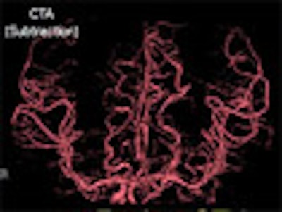
Another promising clinical application is brain imaging of patients suspected of stroke, Katada said. One programmed scan sequence could be used to collect the entire study, from noncontrast CT, to contrast-enhanced CT for CT angiography (CTA), to the CT perfusion study. "All these three can be done in a single sequence," Katada said. "Three scans at one time, and that will help reduce the time for the diagnosis. Time is brain."
 |
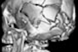
| CT coronary angiography: 64-slice versus 256-slice. |
A third promising application is CT digital subtraction angiography, which Katada believes will be particularly well-suited to area-detector CT as the studies can be performed without moving the patient, leading to greater accuracy in matching subtraction images.
 |
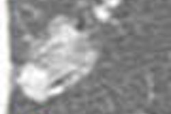
| CTA and perfusion CT images of a left internal carotid artery occlusion. |
Addressing dual-source CT
One of the hottest topics at the MDCT meeting was dual-source CT, which uses two sets of x-ray tubes and detector arrays housed in a single CT gantry. Siemens Medical Solutions of Malvern, PA, introduced its dual-source CT system at the 2005 RSNA meeting and currently has the only dual-source system on the market. Several presentations at the San Francisco meeting described clinical experiences with the technology.
Katada does not see 256-slice CT and dual-source CT as being mutually exclusive. He views dual-source CT as a technique to enhance rotation speed without increasing a scanner's true rotation speed to gain very high temporal resolution. Meanwhile, area-detector scanning as exemplified by 256-slice CT enhances the speed of volume acquisition and coverage.
Using dual-source CT with scanners that have 64 slices or fewer still requires users to work within the helical-scanning paradigm, he said. The required movement of the patient couch with helical dual-source scanning may also impact the accuracy of subtraction imaging with dual-energy x-ray sources (one of the potential new clinical applications possible with dual-source CT) by making it more difficult to match coordinates perfectly, he believes.
It is theoretically possible to merge multiple-source technology with area-detector CT, resulting in a scanner that has both very high temporal resolution and wide anatomical coverage, Katada said. "There is no limitation in merging these technologies, so why don't you combine all of them?" he said. "I always say that the explosion of CT techniques has only just begun."
By Brian Casey
AuntMinnie.com staff writer
August 18, 2006
Related Reading
Dual-source imaging promises better CT scanning, June 15, 2006
Prototype 256-slice CT scanner creates high-res images in a heartbeat, March 14, 2006
Motion-free heart images revealed with 256-slice CT, October 24, 2005
Copyright © 2006 AuntMinnie.com