A 4D ultrasound technique can create "digital casts" of the fetal cardiac chambers and great vessels, opening the door for expanded detection of congenital heart disease, according to an article in the April issue of the Journal of Ultrasound in Medicine.
"The application of spatiotemporal image correlation, inversion mode, and B-flow imaging generates information about the anatomy and pathologic characteristics of the fetal heart (digital casts) that cannot be obtained with 2D fetal echocardiography," wrote a study team led by researchers from the National Institutes of Health. "We propose that these modalities enhance the information provided by ultrasonographic interrogation of the fetal heart and will improve prenatal diagnosis."
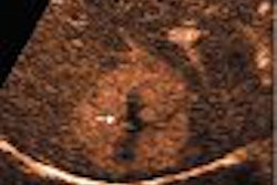
The research team employed spatiotemporal image correlation and inversion mode to examine volume datasets of 23 fetuses with confirmed congenital anomalies (JUM, April 2005, Vol. 24:4, pp.415-424). A subset of studies was also examined with B-flow imaging, and digital reconstructions from abnormal hearts were compared with a library obtained from fetuses without abnormalities.
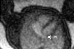
Produced using inversion mode, rendered images of the four-chamber view were characterized by echogenic chambers; sharp delineation of chamber contours when compared with 2D images; and distinct display of the myocardium, interventricular septum, interatrial septum, and mitral and tricuspid valves as anechoic structures, according to the researchers. Ventricular septal defects (VSDs), abnormal differential insertion of the atrioventricular valves, and valve atresia were also visualized well.
As a result, the study team suggests incorporating inversion mode into prenatal diagnosis of congenital heart disease.
"Thick-slice rendering of the four-chamber view with the inversion mode led to improved definition of the cardiac chambers and atrioventricular valves when compared with the corresponding 2D images," the authors wrote. "This feature may be useful in the detection of VSDs, especially in cases in which the original 2D images are of poor quality."
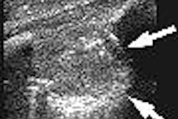
The researchers also stated that they found that 4D reconstruction of the outflow tracts with either inversion-mode or B-flow imaging produces images analogous to those obtained postmortem by injecting silicone rubber to create casts of the cardiovascular cavities.
"Digital casts created with inversion-mode or B-flow imaging allowed visualization of the relationships, size, and course of the outflow tracts in several cardiac anomalies (coarctation of the aorta, pulmonary stenosis, pulmonary atresia, double-outlet right ventricle, and transposition of the great arteries)," the team wrote.
Examiners may also be able to better understand the spatial relationships between the vessels in cases of congenital heart disease, according to the authors. In addition, the combination of inversion-mode and B-flow imaging may help explain cardiac anomalies to parents, particularly for complex malformations.
The authors cautioned, however, that training will be required before these techniques can be introduced, as the technology for reconstructing the outflow tracts of the fetal heart by 4D ultrasound using either inversion-mode or B-flow imaging is relatively new.
Study limitations included knowledge by the examiner of the final diagnosis before reconstructing the volume datasets, as well as not enough examples of cardiovascular anomalies with a confirmed postnatal outcome to compare 3D reconstructions produced by inversion-mode or B-flow imaging, the authors stated.
"Although both techniques are capable of producing digital casts of cardiovascular cavities and blood vessels, B-flow imaging may be able to provide better border definition and fewer artifacts than the inversion mode," they wrote. "B-flow technology digitally enhances signals from weak blood reflectors from vessels and, at the same time, suppresses strong signals from surrounding tissues."
By Erik L. Ridley
AuntMinnie.com staff writer
April 6, 2005
Related Reading
3D volume US shows promise for second-trimester fetal evaluation, March 17, 2005
3D ultrasound poised to revolutionize pelvic imaging, September 9, 2004
3D and the future of imaging, November 3, 2003
3D ultrasound offers reliable assessment of gallbladder, bile ducts, May 5, 2003
Copyright © 2005 AuntMinnie.com