Computer-aided detection (CAD) technology can yield sensitivity gains for all pulmonary nodule types detected on CT scans, according to research presented at the recent American Roentgen Ray Society (ARRS) 2009 annual meeting in Boston.
"The use of CAD as a second reader significantly improves radiologists' detection of both subsolid and solid nodules," said Dr. Myrna Godoy of New York University Langone Medical Center in New York City. She presented the findings during a scientific session at the show.
Although the use of CAD systems as a second reader has been shown to increase detection of pulmonary nodules, current systems are designed primarily for detecting solid nodules. However, subsolid nodules are also clinically important, as ground-glass nodules and part-solid nodules often indicate adenocarcinoma and its premalignant forms, according to Godoy.
The research team sought to evaluate a CAD prototype (Siemens Healthcare, Malvern, PA) designed for improved performance in detecting subsolid nodules. The group wanted to assess the systems' value for detecting both subsolid and solid pulmonary nodules on CT scans, and also to evaluate the technology's effect on radiologists' detection of pulmonary nodules.
Four chest radiologists independently reviewed 1.0-mm sections from 46 chest MDCT scans with known subsolid nodules on a DICOM viewer. Cases were selected from a pool of cases with lung nodules from two institutions and were not previously used for CAD development or training of the system. Each radiologist marked all lung nodules and rated confidence as either less than 50% or greater than 50%.
The nodules were characterized by maximum diameter and attenuation, which included pure ground-glass, part-solid, and solid nodules, Godoy said. A fifth experienced radiologist resolved any differences between readers for the presence or characterization of the nodules and provided the ground truth for the study.
On the 46 scans, 270 nodules were adjudicated as ground truth. Nodules less than 4 mm and ground-glass nodules less than 6 mm were excluded, leaving 118 nodules for inclusion in the study.
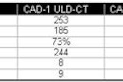
These nodules had a median maximum diameter of 5.5 mm (range, 4.0-27.5 mm) and included 62 solid nodules, 20 part-solid nodules, and 36 pure ground-glass nodules. The prototype CAD algorithm was then applied to the CT scans. CAD yielded 79% sensitivity for all nodules, 87% sensitivity for solid nodules, 85% for part-solid nodules, and 61% for ground-glass nodules.
CAD significantly increased reader sensitivity in all subgroups (p < 0.001), according to Godoy.
Reader sensitivity improved from a mean of 68% before CAD for all nodules to 83% after CAD. For solid nodules, mean sensitivity increased from 60% to 85% for solid nodules. Mean sensitivity also climbed from 80% to 95% for part-solid nodules and from 75% to 86% for ground-glass nodules.
As for false positives, CAD produced a false-positive rate ranging from zero to 13 per scan, with a mean of 3.4 per scan. Readers had an average false-positive rate of one per scan, with a range of zero to six. The use of CAD did not increase the number of false positives for any of the readers, she said.
By Erik L. Ridley
AuntMinnie.com staff writer
May 15, 2009
Related Reading
Computer-aided system increases detection of early-stage lung cancer, May 4, 2009
Integrating lung CAD with PACS boosts utilization, March 6, 2009
CAD helps novice readers detect pulmonary embolism in CT studies, February 12, 2009
Lung CAD shows promise as concurrent reader, January 30, 2009
CAD may boost lymph node detection, December 4, 2008
Copyright © 2009 AuntMinnie.com