Joint Photographic Experts Group (JPEG) 2000 compression produces more artifacts in thin-section lung CT images than in thick-section images, according to research published in the American Journal of Roentgenology.
"Section thickness should be taken into account when adjusting the compression level for lung CT images," wrote a research team from Seoul National University Bundang Hospital in Korea.
To determine the differences in artifacts between thin- and thick-section lung CT images, the Korean researchers compressed 35 thin-section (1 mm) and 35 corresponding thick-section (5 mm) images using PICTools JPEG 2000 compression software (Pegasus Imaging, Tampa, FL). In each case, pixels outside of the lung were replaced with those from the original image (AJR, August 2008, Vol. 191:2, pp. W38-W43).
Three radiologists then independently rated the compression artifacts, providing grades of 0 (indistinguishable), 1 (barely perceptible image differences), 2 (subtle image differences), or 3 (significant image differences). The researchers also compared the thin and thick sections at each compression level for peak signal-to-noise (S/N) ratio.
The study team found that thin sections had a smaller peak S/N ratio (p < 0.0001) and a higher grade of artifacts than thick sections. Significant differences were shown at compression levels of 10:1 (mean score of 0.8 versus 0.4, 0.9 versus 0.1, and 0.3 versus 0.0; p < 0.009 for the three readers) and 15:1 (1.9 versus 1.0, 1.9 versus 1.1, 1.5 versus 0.6; p < 0.0001).
The researchers also noted that the percentages of distinguishable pairs (grades 1-3) were greater for thin sections, with a statistically significant difference at 10:1 for two readers (31% versus 3% and 74% versus 37%; p < 0.006).
The authors acknowledged several limitations of the study, including an inability to completely blind the readers to whether the tested image was a thin or thick section, the use of images with relatively homogenous scanning and reconstruction parameters, and a limited number of images.
"The lung shows more JPEG 2000 compression artifacts on thin-section CT images at a lung window setting than on thick-section images," the authors concluded.
Radiologist perception
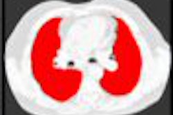
In a related study also published online in the American Journal of Roentgenology, additional research led by the Seoul National University Bundang Hospital found regional differences among radiologists in their perception of artifacts in low-dose chest CT images.
"Although JPEG 2000 compression introduces greater mathematic artifacts in the lungs than in the chest wall and mediastinum in low-dose chest CT images with a lung window setting, radiologists perceive fewer artifacts in the lungs than in the chest wall and mediastinum," the authors wrote.
The study team sought to investigate the differences of perceptible artifacts and to show that the high-dynamic range visual difference predictor (HDR-VDP) perceptual image quality metric can reproduce regional differences in artifacts (AJR, August 2008, Vol. 191:2, pp. W30-W37).
The researchers compressed 20 images reversibly and irreversibly up to 6:1-30:1 using PICTools (Pegasus Imaging). Compressed pixels outside of each tested region were replaced with the original pixels.
Three radiologists then independently rated the compression artifacts as grade 0 (none, indistinguishable), grade 1 (barely perceptible), grade 2 (subtle), or grade 3 (significant). The two regions (lungs; chest wall and mediastinum) were then compared at each compression level for the readers' responses, peak S/N ratio, and HDR-VDP results.
The researchers found that readers rated artifacts as lower grades in the lungs than in the chest wall and mediastinum, with statistically significant differences at 10:1-20:1 for reader 1, 8:1-15:1 for reader 2, and 8:1-20:1 for reader 3. The grade of zero (no artifacts, indistinguishable) was more frequently given in the lungs, reaching statistical significance at 10:1 for reader 1 and 8:1-10:1 for readers 2 and 3.
The peak S/N ratio results also indicated greater artifacts in the lungs (p < 0.001), while the HDR-VDP results found fewer lung artifacts (p < 0.001).
"The tested perceptual image quality metric (HDR-VDP) can successfully reproduce such a regional difference in radiologists' perceptions of the compression artifacts," the authors concluded.
By Erik L. Ridley
AuntMinnie.com staff writer
August 13, 2008
Related Reading
Lossy compression perceptible at even low levels, May 15, 2008
Lossy compression performs well in abdominal CT images, January 13, 2008
Canadian research finds no loss with lossy compression, August 14, 2007
Enhanced DICOM objects, compression help with MDCT, MR challenges, March 23, 2007
Lossy 3D JPEG 2000 compression useful for abdominal MDCT, February 23, 2007
Copyright © 2008 AuntMinnie.com