Computer-aided detection (CAD) technology can assist in diagnosing pneumonia on chest radiographs in children, according to research published in the August issue of the International Journal of Medical Informatics.
"[The] Pneumo-CAD system could represent a complementary tool in monitoring the burden of bacterial pneumonia at the pediatric population level to support the public health authorities in the decision of introducing the vaccination against pneumonia," wrote a Brazilian research team led by Leandro Luís Galdino Oliveira of Universidade Federal de Goiás in Goiânia, Brazil.
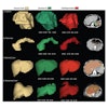
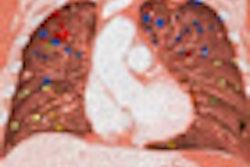
The Brazilian researchers developed Pneumo-CAD, a CAD prototype designed to be able to discriminate chest radiological patterns between pneumonia of bacterial etiology from absence of pneumonia in a population of patients previously evaluated by an attendant pediatrician. The system employs wavelet transforms to extract lung image features and a weighted "nearest-neighbor" based on a Euclidean metric to measure the similarity between images (Int J Med Inform, Vol. 77:8, pp. 555-564).
The researchers tested the system, using eight different wavelet transforms, on a total of 1,000 x-ray images from a pneumonia surveillance database. The radiographs had been captured using a digital camera at a resolution of 1024 x 768 pixels, with 8-bit grayscale. Images were concurrently read by two independent radiologists for the presence of pneumonia.
The radiologists had confirmed the diagnosis of pneumonia in 74% of the cases, found a normal chest radiograph in 22% of the cases, and confirmed other diagnoses in 4% of the cases. For the purposes of the study, these diagnoses were considered to be the gold standard.
The researchers sequentially retrieved a subset of 40 cases (20 normal, 20 with a diagnosis of pneumonia) to build up the knowledge base of the Pneumo-CAD system. An additional 20 cases (10 normal, 10 with diagnosis of pneumonia) were randomly selected to evaluate the system's image classification performance.
The researchers then tested the system with eight different wavelet transforms using two methodologies. The first methodology worked with a rank-weighted 15-nearest-neighbor scheme, while the second one employed a distance-dependent weighting for image classification, according to the researchers.
The study team found that the Pneumo-CAD system produced the best accuracy when using the Haar wavelet transform, yielding an area under the ROC (receiver operator characteristics) curve of 0.97 for methodology I and 0.94 for methodology II in discriminating between normal and pneumonia cases. Sensitivity was 100% for both methodologies, while specificity was 80% for methodology I and 90% for methodology II.
"Thus, confronting the results of both methodologies, we found that methodology I, which used the 15 most similar images out of the 40 images in the knowledge database, proved to be more capable of correctly classifying chest radiographs with suspicious diagnosis of pneumonia," the authors wrote.
Applying such computerized tools could standardize the interpretation of chest radiographs and increase the probability that any difference between studies reflects the real geographic differences of pneumonia epidemiology and differences in vaccination effectiveness, the authors concluded.
By Erik L. Ridley
AuntMinnie.com staff writer
July 23, 2008
Related Reading
CAD software finds vertebral fractures in digital chest exams, July 7, 2008
CAD helps residents identify lung nodules on DR, May 20, 2008
Criteria determine CAD mark sensitivity, March 20, 2008
CAD provides mixed benefits for DR lung exams, March 8, 2008
DR image processing produces mixed CAD results, February 12, 2008
Copyright © 2008 AuntMinnie.com