The future can't come fast enough for some advanced imaging enthusiasts. There is the long, sometimes painfully slow disconnect between the development of a promising algorithm or software function and its delivery as a real clinical tool to aid diagnosis and therapy.
Systems that can perform great feats in more than one anatomic region or in multiple diagnostic settings are even rarer. But for Kensaku Mori, Ph.D., and colleagues at the Graduate School of Information Science at Nagoya University in Japan, that kind of gee-whiz future is approaching fast.
To be sure, the NavI-CAD (navigation-based intelligent computer-aided diagnosis) system Mori discussed at the 2008 Computer Assisted Radiology and Surgery (CARS) meeting in Barcelona, Spain, is a work in progress. Some components aren't ready for prime time. Other features can be found in somewhat similar form elsewhere. And in any case, you can't put your hands on a NavI-CAD unless you happen to work in one of a handful of Japanese research hospitals. But a decade of research on the group's landmark project, supported in part by the Japanese government, has yielded a pretty useful set of advanced imaging tools.
Why do we need navigation-based CAD? Recent years have produced remarkable progress in the field of volumetric imaging, with multidetector-row CT and high-field MRI, but we still lack highly functional ways to navigate the human body and use the images these machines were designed to produce, Mori said in his CARS presentation.
The navigation-based intelligent CAD system fuses computer-aided diagnosis and navigation-based diagnosis, enabling doctors to navigate inside a human body, he said.
"We need efficient and effective assistance for reading such images for computer-aided diagnosis and computer-assisted surgery," Mori said. The NavI-CAD "assists and augments the diagnostic process by employing advanced visualization techniques."
Navigation-based diagnosis refers to exploration inside the virtual human body, including real-time visualization, advanced human interface, and augmented image displays based on computer analysis, Mori explained. Data from human patients are processed and ready for exploration in a few minutes. Fly through a region and get measurements, lesion locations, and path information.
Poke a virtual organ and you can move it, turn it around, and inspect the other side. You'll have only the patient data you acquired, so organ interaction doesn't mean you should expect the kind of advanced, haptically driven soft-tissue deformations researchers have begun to incorporate into surgical training modules. But you can visualize CT data in many intuitive and flexible ways.
"The target of navigation-based diagnosis consists of two things: one, navigation in physical space, and two, navigation in logical space," Mori said. The physical space includes the space axis (position, orientation), the time axis (time of acquisition), the transfer function of the intensity axis, and the scale axis of image resolution.
The other side of the coin, the logical or meaning space, enables observation of organs in a defined order, observation along with the anatomical structure (lesion axis), and observation of lesions detected automatically. "Navigation in logical space is quite important to the computer-aided diagnostician," Mori said.
Chest
The logical function of anatomic structure analysis in the chest, for example, can be used to segment the bronchus, classify pulmonary arteries and veins, and provide the nomenclature for each branch and vessel segment that pops up during a fly-through, as Mori demonstrated. This feature has implications for faster radiology reporting.
 |
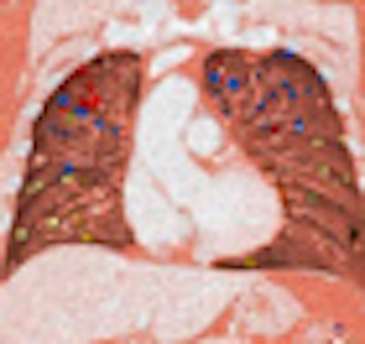
| NavI-CAD performs lung navigation, lobe segmentation, nodule detection, and other functions. All images courtesy of Kensaku Mori, Ph.D. |
Turn on the lung CAD function and solitary pulmonary nodule candidates are highlighted. NavI-CAD can measure volume, cross-sectional areas, and suspicious lesions along with the names of the vessels to which they're attached. Another function divides the organ into lung lobe regions, with fissure extraction based on a Hessian matrix algorithm, quadratic curve-fitting, and division of lung lobes.
 |
| Segmental lung lobe division functions are based on 3D image decomposition. |
A figure decomposition technique is used to extract the lung lobe regions from 3D chest CT images, Mori explained. Navigation through the airways is facilitated by anatomic name navigation as well as a 3D virtual bronchoscopy fly-through. The lung parenchyma can even be examined dynamically via time-lapse visualization from CT data acquired in two phases. The lung lobe division functionality can also erode 3D images, removing small connected components and dilating larger connected components to the appropriate lung region.
 |
| Navigation to lung nodules is performed by fly-through as well as anatomic name navigation, a functionality that will be expanded to other organ systems and anatomic levels in future versions of the system. |
At this year's CARS conference, Mori also presented a study on fiducial-free bronchoscope tracking based on nonrigid transformation methods.
Colon
The presence of haustral folds can cause distortion in volume rendering of virtual colonoscopy data, so NavI-CAD employs a circular ray-casting method based on a spring model to reduce this distortion, Mori said.
For virtual colonoscopy exams, the navigation-based CAD system provides a synchronized display of five colon views. Automated polyp detection is achieved with local intensity structure analysis from 3D CT data.
 |
| The system displays up to five synchronized views, a new feature in virtual colonoscopy imaging. |
The recently updated colon analysis function includes smoothing by Gaussian filter, detection of polyp candidates, and enhancement of blob structures by eignenvalues of a Hessian matrix, Mori said. Subsequent steps include false-positive reduction, connection of low-output components, and elimination of small connected components. (A 2006 version of the colon navigation and CAD system was previously featured on AuntMinnie.com.)
 |
| Polyp detection feature marks true-positive lesions of the colon. |
Laparoscopy
"CAD can also be useful in assisting surgery," he said. "The surgeon needs high skill levels for laparoscopic surgery," due to the narrow viewing area of the endoscope. "We need some techniques to make it much safer," Mori said.
From preoperative CT data, the NavI-CAD extracts and segments the abdominal organs based on two 3D CT images from different phases, registering them with nonrigid image registration. The blood vessel network and lymph nodes are depicted, and the virtual laparoscope with virtual wall lift can be used for surgical planning. Color coding of the organs demonstrates the precise reach of the laparoscope.
 |
| Color coding of target organs demonstrates the precise reach of the laparoscope for safer surgical navigation. |
The road ahead
Mori's future goals for the system are ambitious. In its current form, NavI-CAD uses only local anatomic information, for example, but whole-body anatomic information is needed for advanced CAD and computer-assisted surgery systems, he said. More level-of-detail information is needed top to bottom, with labeling throughout the human body spanning the range from macro to nano levels, Mori added.
NavI-CAD needs to reflect all medical stages from diagnosis to treatment, incorporating intelligent media in the labeling development, Mori said. Patient data are not yet captured in the operating room. DICOM compliance is essential at all levels.
"Also, we need real-time CAD for the operating room," he said.
By Eric Barnes
AuntMinnie.com staff writer
July 7, 2008
Related Reading
CARS report: Fuzzy theory yields sharper CTA images, June 27, 2008
CARS report: Imaging workstations key to patient-specific care, June 27, 2008
Automated polyp measurement cuts variability, May 21, 2008
Part III: Image processing has room to grow, October 23, 2006
VC visualization tool combines advanced features, August 17, 2006
Copyright © 2008 AuntMinnie.com