ATLANTIC CITY - Volume sonography yields efficiency and workflow improvements, and can be the technology ultrasound needs to regain market share from other cross-sectional imaging applications, according to Dr. Beryl Benacerraf of Harvard Medical School in Boston.
"We need to move ultrasound into applications and efficiency previously inaccessible to us, but available to CT and MR," Benacerraf said. She spoke during a talk at this week's Leading Edge in Diagnostic Ultrasound conference.
In a research project, Benacerraf and Harvard researchers sought to determine whether volume sonography can be a quicker and more efficient screening exam of the second-trimester fetus. They also wanted to see whether the fetus could be "put through" the 3D ultrasound scanner, much like a child or adult goes through a CT scanner, she said. In addition, they aimed to find whether the entire exam dataset could be sent automatically to a central reading area for later review.
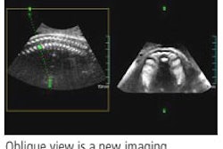
The study included 50 consecutive 17- to 21-week fetuses that were scanned for a structural survey with a normal 2D scan. After the standard 2D sonograph was performed by one of eight sonographers, the sonographers then obtained volume acquisitions in the regions of the head (transverse), thorax (transverse), face (longitudinal), and lower extremities (longitudinal).
At least one to two weeks after the initial scans, the volumes were reviewed on unmarked CDs by three physicians on a separate workstation. The researchers then calculated the time used to obtain the volumes, the time used to do the fetal surveys on the five volumes including biparietal diameter (BPD) and femur measurements, the completeness of structural survey, and the time needed to complete the standard 2D study.
The 3D volume findings were then compared to the 2D exam to determine whether the 3D offline review was any less accurate.
The frequency of identifying all anatomic landmarks on 3D was at least 94%, with the exception of both fetal arms (92%). Overall, 3D BPD and femur measurements were also highly congruent with 2D measurements (within 1 mm on each side), and differences were not judged to be clinically significant, Benacerraf said.
In time comparisons, obtaining the volumes took a mean time of 1.8 minutes, with a mean time to do reconstruction ranging from 4.8 to 5.5 minutes, for a total mean time for the entire volume scan ranging from 6.6 to 7.3 minutes, she said. The 2D surveys took a mean time of 19.4 minutes.
As a result, the volume scans saved 12 to 13 minutes of machine/sonographer time per patient, with total room time-savings for the 50 patients of 10.3 to 10.8 hours. These findings suggest that 3D offers the opportunity for ultrasound practices to dramatically save time, Benacerraf said.
"We could be on the threshold of improving the efficiency and workflow of our ultrasound units," she said.
3D issues
The practice of 3D sonography comes with its own issues, including patient perception and satisfaction with a much shorter exam time. Patients may not like being on the table for just two minutes for an obstetrical ultrasound exam, Benacerraf said.
"Patients may need to be educated that what comprises the medical part of the ultrasound exam," she said. "You can't lengthen the study just to please the patient. Certainly, there is no other modality that lengthens or prolongs the exam beyond what is really medically necessary or indicated just to please the patient."
As for concerns over incomplete exams, a couple of additional images will be necessary to complete the scan, such as a picture of the placenta, cervix, and a video clip of the beating heart. This might add less than 30 seconds to the screening scan, Benacerraf said.
To be complete, 5% to 10% of the scans may require additional views in 2D traditional scanning, she said.
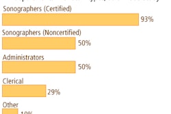
Adoption of 3D exams will also impact sonographers, Benacerraf said. Images will not be as operator-dependent, and sonographers will spend less time scanning. That may improve the repetitive injury problems they have, she said.
"I have 11 sonographers that work for me, and they are very excited about this," Benacerraf said. "Many of them have been doing sonography for a long, long time and have a lot of orthopedic problems that stem from scanning for a long, long time. And they love 3D."
In addition, sonographers may well be called upon to reconstruct views offline from the volumes, a development that will vary their tasks and average physical time, she said.
"As (3D sonography) starts to evolve, (the sonographer) will probably take on multiple roles," Benacerraf said. "This is going to vary their job description and, in my opinion, reduce their injuries. This should not be a threat at all to sonographers, but more of an opportunity to do lots of different things."
Three-dimensional sonography will also benefit physicians, she said. For physicians that scan the patient, the time for the virtual scan is probably an improvement over walking into the room, meeting the patient, and doing the scan live, according to Benacerraf.
And for those who review sonographers' images, the sonographer can perform the 3D image manipulations after obtaining the volumes, and display the images not unlike the usual image displays, Benacerraf noted. Other advantages include the ability to generate missing images offline by "virtual scanning" even when the patient is gone, she said.
With the advent of 3D, the ultrasound community must now discover the areas that have become accessible to the modality due to the ability to reconstruct any plane and scan it in real-time, Benacerraf said.
"CT and MR have really taken the market share that ultrasound used to have," she said. "I think it's time for us to regain some of our market share, and it's really up to us to discover how to move this forward and make this 3D concept reality for those of us that practice ultrasound. And I think this progress will allow us to maintain and cement ultrasound's role in cross-sectional imaging applications."
By Erik L. Ridley
AuntMinnie.com staff writer
May 25, 2006
Related Reading
3D sonography of the endometrium adds value, May 5, 2006
3D ultrasound predicts fetal pulmonary hypoplasia, April 21, 2006
3D ultrasound shows value in detecting fetal malformations, March 24, 2006
3D imaging creates paradox for ultrasound vendors, November 9, 2005
Practical tips ease 3D/4D ultrasound, August 12, 2005
Copyright © 2006 AuntMinnie.com