Lung volume measurements from 3D ultrasound studies appear to aid in diagnosing fetal pulmonary hypoplasia, according to researchers from the VU University Medical Center in Amsterdam, Netherlands.
To study the potential of using lung volume measurements on 3D ultrasound in predicting pulmonary hypoplasia in complicated pregnancies, the VU researchers prospectively studied 51 patients. All 51 pregnancies (with a gestational age of 18-36 weeks) were complicated by various disorders or had complications with regard to pulmonary hypoplasia, according to Dr. Franca Gerards. She presented the research in March at the annual convention of the American Institute of Ultrasound in Medicine (AIUM) in Washington, DC.
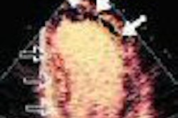
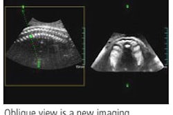
One to four scans of the fetal lung were obtained. The researchers acquired two to five volumes of the entire chest, using the freehand with positioning method, Gerards said.
An examiner then manually traced each lung in the transverse plane from the fetal clavicles to the dome of the diaphragm and measured three times. Using these 2D outlines, the ultrasound scanner's built-in program automatically constructed a 3D model of the lung, according to the researchers.
The lung volume measurements were then compared with the longitudinal reference intervals of fetal lung volumes for each gestational age obtained from uncomplicated pregnancies.
Complications included preterm rupture of the membranes (18), renal and urinary tract anomalies (11), skeletal and neuromuscular pathologies (8), and other conditions (6).
Fourteen (27.5%) of the 51 infants were diagnosed with pulmonary hypoplasia on postmortem examination, and in eight cases (15.7%), pulmonary hypoplasia was suspected due to clinical and radiologic presentation.
Combining the oligohydramnios caused by the premature rupture of membranes, the renal and urinary tract anomalies, the skeletal and neuromuscular malformation, and other disorders suspected for pulmonary hypoplasia based on gestational age yielded overall sensitivity of 86% (confidence interval ± 15%), specificity of 100%, positive predictive value of 100%, and negative predictive value of 88% (confidence index ± 12.7%) in predicting pulmonary hypoplasia.
Comparing lung volume measurements of the intrauterine growth-restricted fetuses caused by uteroplacental insufficiency with the normal curves based on estimated fetal weight resulted, however, in 100% sensitivity, specificity, positive predictive value, and negative predictive value. The intraclass correlation coefficient was 0.99 for both lungs.
"3D lung volume measurements seem to be useful in predicting pulmonary hypoplasia prenatally," Gerards said.
By Erik L. Ridley
AuntMinnie.com staff writer
April 21, 2006
Related Reading
Ultrasound alters management of infants with genitourinary disorders, October 14, 2004
Embryo and Fetal Pathology: Color Atlas with Ultrasound Correlation, October 13, 2004
Three-dimensional ultrasound shows utility in fetal lung volume measurements, March 18, 2004
Part II: The psychological impact of 3-D ultrasound on pregnant women, December 30, 2003
Part I: The psychological impact of 3-D ultrasound on pregnant women, December 29, 2003
Copyright © 2006 AuntMinnie.com