Last year's Digital Mammographic Imaging Screening Trial (DMIST) pretty much laid to rest doubts about the clinical performance of full-field digital mammography. But a host of questions remain for breast imaging centers thinking of making the switch, ranging from FFDM's impact on workflow to how to handle and store all those digital images.
The benefits of digital mammography are legion, according to Bonnie Rush, president of Breast Imaging Specialists, a San Diego-based consulting firm. Digital mammography offers a dynamic range that film-based mammography can't, allowing radiologists to manipulate images and examine areas in the breast in ways that aren't possible with analog images.
Digital mammography also eliminates chemical processing and its associated personnel expenses (no more trudging to the process room) and external noise factors such as dust; it can also reduce the amount of hard-copy storage a facility needs, and, with a good PACS network in place, ensure that images aren't lost or misplaced. If staff expense is a concern, FFDM can reduce the need for darkroom assistants, radiology aides, chart room staff, and technologists.
Installing a digital mammography system can also reduce recall rates in the long run, according to Rush. "Initially, recall rates and interpretation time increase with digital, depending on how much training radiologists have in the technology prior to the transition," Rush said. "There's a learning curve, sure, but as radiologists get used to looking at digital images, and as they get used to the workstation's tools at their disposal, recall rates will settle down and interpretation time will decrease."
All these technical advantages go hand-in-hand with patient-care benefits, not the least of which is reducing a woman's anxiety during her mammogram. With digital mammography, the technologist never needs to leave the room, and can show the images to the patient right as they're taken, explaining why further images may be needed, Rush said.
"In an analog environment, the technologist leaves to take the film to processing, and while he or she is gone the woman has the opportunity to worry about problems with her mammogram," she said. "Anecdotally, the digital environment helps ease patient anxiety since the technologist stays in the room until the exam is completed and the patient is released."
Finally, digital mammography brings a certain cachet that can boost a facility's market share, both through patient word of mouth and the facility's advertising efforts. This effect is becoming more common as many women are beginning to actively seek out centers with digital technology due to its proven advantages in imaging dense breast tissue.
Installation considerations
So, the verdict is in on FFDM's value. But there are still some challenges to installing digital mammography, especially from scratch. FFDM systems are sold at a premium to analog systems, with a flat-panel digital radiography-based FFDM unit's list price at more than $400,000, and its upkeep can be as much as $135,000 per year. Thus, a facility's return on investment, depending on the number of patients it serves and its particular insurance mix, can take a number of years.
Facilities can expect decreased productivity for a time while imaging staff learns to use the technology; it may be necessary to rearrange personnel or create temporary positions to smooth workflow, according to the 2006 "Mammography Regulation and Reimbursement Report," published by consulting firm HC Pro of Marblehead, MA.
"Since the industry indicates that one DR mammo unit can replace up to two analog units and still maintain current productivity, for facilities with older units, sufficient capital, and a patient base that can support DR, going that direction makes sense," Rush said.
Evaluating patient load and types of insurance within that patient base is crucial to determining whether digital mammography will work. The general rule of thumb is that a facility needs to perform between 15,000 to 25,000 mammography exams per year for a timely return on investment, although these numbers are not gospel, Rush said.
Something else to consider is that going digital paves the way to other services -- such as telemammography or computer-aided detection (CAD) -- that might be impossible or more difficult to conduct without an FFDM system. In some cases, these additional services can mean the difference between treading water and making a profit. For example, adding CAD to FFDM can increase a facility's bottom line without increasing staff cost, and more and more insurance companies are looking at CAD as standard of care for a screening mammogram.
"The question of how many patients it takes per year to make a return on investment is subjective," Rush said. "It's important that a facility review its current patient base, its growth potential, the patient-specific reimbursement rates, and any ancillary services it can offer that will bring additional monies into the department. This last point is particularly important for independent imaging centers not associated with a hospital."
Also crucial to the digital mammography decision is evaluating the facility's existing PACS, or investigating PACS options if no system is in place. Having a powerful PACS that can handle multiple modalities and workstations is key to the success of the transition to digital mammography, according to Gerald Kolb, chief development officer for Solis Women's Health, headquartered in Austin, TX.
Solis currently owns five breast imaging centers in the Dallas-Ft. Worth metropolitan area, is opening two more this month, and is actively acquiring another two. Solis facilities are in the process of converting to digital mammography via computed radiography (CR), installing 17 acquisition/image processing units, together with eight workstations, and all the associated infrastructure, including a fully integrated information management system; they perform more than 85,000 mammography exams per year.
"The fact is you have to be able to work flexibly, with all vendors and modalities," Kolb said. "We want to be prepared in the future to move images around between centers, and to access offsite images -- radiologists want to pull down images that are on other PACS at hospitals and other facilities -- so we've put together a PACS infrastructure that can allow us to be extremely flexible."
The more pixels, the more storage and bandwidth are necessary, and facilities can expect each unit to produce 1 to 3 terabytes of information per year. Hiring a PACS administrator (and deciding whether that person is clinically or technologically based) can be helpful as well, according to HC Pro. Kolb brought all the vendors and staff involved in the transition into weekly meetings to decrease the chance of miscommunication.
The experience at Solis brings up a good point -- facilities going digital also now have a choice of technologies, a choice that didn't exist before 2006, when the U.S. Food and Drug Administration approved the first digital mammography system based on CR. The technology enables facilities to go digital with their existing mammography equipment (although they would need to install a CR reader if they didn't already have one).
Choosing between flat-panel DR or storage-phosphor CR depends on a number of factors, including the size of the facility, its patient load, whether it has more up-front capital or less, and the age of its existing equipment, according to Rush. For a facility already invested in CR, or with a smaller patient volume and fewer opportunities to boost referrals, CR makes sense, especially since the CR mammography readers can also do general x-ray exams.
Finally, before making any decisions, schedule a number of site visits, according to Rush.
"Once you choose a vendor, insist on going on site visits," she said. "For a smooth transition, it's important that all those involved in supporting, implementing, and using the new digital system fully understand the impact of the changeover."
The IT side of digital mammography
BreastScreen Waitemata Northland (BSWN) in New Zealand was established in February 2006 and serves a population of 93,000, performing about 24,500 exams annually. The two main sites in Takapuna, Auckland, and in Whangarei are both fully digital; a third digital site is planned to open in July of this year. Since the two sites were new, there was no transition from analog to digital. Instead, the focus was on analyzing the cost benefits between the two modalities, according to Andrew Cave, business project manager. BSWN was able to offset costs such as building in a film processor against the cost of setting up a digital site.
"Teleradiology was a key consideration," Cave said. "Our two digital sites are 250 kilometers apart, and the northern site has only two radiologists. One of our clinical requirements is that no two radiologists should consistently perform blind reads against each other. Digital mammography allowed us to meet this requirement without having the double handling involved with loading a viewer at one site for the first read, pulling the files down, transferring them, and then reloading another viewer for the second read."
BSWN is operated as part of the Waitemata District Health Board, and because of this, existing PACS and RIS networks were in place at the time the two digital mammography breast imaging sites were established. But these existing systems were inadequate to support the specific requirements of digital mammography.
In New Zealand, all mammograms performed under the national breast screening program (BreastScreen Aotearoa) are read independently by two radiologists, with discordant opinions resolved by a third read, consensus, or arbitration. To ensure that this requirement was adequately met, BSWN built in additional functionality into its breast screening information system and installed a dedicated PACS for breast imaging. To cope with the extra storage demands, BSWN uses a dedicated archive with setup costs split among the cardiology, radiology, and breast screening departments.
"One thing we learned early on was that we needed to have the image cache right next to the reporting workstation," Cave said. "Despite being connected to a 100-Mbps link, high network latency was adding up to 30 seconds reading time for each study."
BSWN changed its network architecture so that all sites where readings are done have access to the image cache. Radiologists' workstations and the image cache now sit on a 1-Gbps network, with no routers involved.
"This change gives good performance -- our radiologists are happy -- and it means we can operate our remote sites over a 10-Mbps link, thus reducing operating costs, which makes our business managers happy," Cave said.
Small centers make the shift
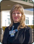
"The exam is quicker, and if the tech does have to repeat an exam, she can explain it to the patient right there."
-- Mary Ann Fitzgerald, Fairchild Medical Center, Yreka, CA
"Geographically, we're a long way from any other hospital, with mountain ranges in between," Fitzgerald said. "We wanted people in our area to have the best technology without having to go elsewhere, and, since the rest of the hospital was already digital, it was a natural move (to do so with mammography)."
Fairchild went digital in August of 2005, and added digital mammography to the mix in May 2006, increasing its PACS storage capability to add the new mammography exams, as well as priors that had been digitized. The biggest benefit Fitzgerald has seen since transitioning to digital mammography is the quality of the images and the patients' comfort level.
"Women aren't left alone in the room anymore," she said. "The exam is quicker, and if the tech does have to repeat an exam, she can explain it to the patient right there."

"We considered replacing our analog unit with another analog, but instead chose to move ahead with digital and have actually increased the number of exams by 10% per day."
-- Paul Conklin, St. Mary Medical Center, Walla Walla, WA
"We considered replacing our analog unit with another analog, but instead chose to move ahead with digital and have actually increased the number of exams by 10% per day," Conklin said.
CAD was important to St. Mary's for its long-term benefit, Conklin said, and the addition of CAD has increased the general sense of confidence in a small mammography department.
"The addition of CAD has definitely reduced the number of overreads we're doing," he said.
Conklin and his staff conducted an analysis on what their return on investment would be going from an analog system that had been completely paid off and had depreciated, and estimated that it will take five years to start making a profit on the new digital machine with current volumes.
Managing the comfort factor
The biggest challenge for St. Mary's was making radiologists comfortable with the switch from rotor image viewing to a digital workstation, according to Conklin. The facility's radiologists underwent an eight-hour training course given by the device vendor.
"Our radiologists were already aware of the benefits and challenges of digital," he said. "In addition to the training, it took about two or three weeks for them to get back to their normal reading speeds," he said.
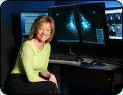
"Going digital offers the possibility of earlier detection, and that's what we're trying to accomplish."
-- Dr. Elizabeth Jekot, Elizabeth Jekot, M.D. Breast Imaging Center, Richardson, TX
"Digital improves throughput by decreasing exam time for the patient and the technologist," Jekot said. "But it does increase read time for the physician, just because there's so much more information. Even with practice and improved hanging protocols, I doubt breast imagers will be as fast at reading digital exams as with film-screen any time soon. But going digital offers the possibility of earlier detection, and that's what we're trying to accomplish."
By Kate Madden Yee
AuntMinnie.com contributing writer
March 1, 2007
Copyright © 2007 AuntMinnie.com


