The potentially detrimental effects of premature birth are both immediate and long-term. While many of these babies do grow up to be perfectly healthy adults, others must contend with the aftermath of a difficult entry into the world.
A recent study by psychiatrists at King's College in London found diffuse white- and gray-matter abnormalities in individuals with very low birth weight. Using functional MRI, they looked at a group of 23-year-old adults, all of whom were born at birth weights below normal. Imaging showed that lateral ventricular volume was significantly increased in these individuals, as was the ratio of gray to white matter. The authors attributed these anomalies to perinatal brain injury (Developmental Medicine and Child Neurology, January 2004, Vol. 46:1, pp. 179-183).
Another study, this time using 11C-raclopride PET, found that one of the environmental factors that contribute to attention deficit/hyperactivity disorder (ADHD) is preterm birth and cerebral ischemia. This can lead to persistent deficient dopaminergic neurotransmission, which is a pathophysiological hallmark of ADHD, explained the authors, from Aarhus University Hospital in Denmark (Developmental Medicine and Child Neurology, March 2004, Vol. 4:3, pp. 179-183).
Both of these investigators successfully used imaging on adults who survived preterm birth; neurological scans performed on infants, while they are still in the danger zone, is much trickier. Preterm babies cannot be moved and require an environment that is carefully controlled. But imaging -- and MRI specifically -- may offer the best way to effectively diagnose certain pathologies in these children.
To that end, radiologists at a U.K. institution have shared their experiences with an MR incubator designed especially for the smallest patients.
MRI incubator
"There have been several attempts to overcome the problem of imaging the neonate, including dedicated scanners on nenonatal intensive care units," wrote Dr. Elspeth Whitby from the section of academic radiology at the University of Sheffield and the Royal Hallamshire Hospital, both in Sheffield. "However, (these scanners) are not widely available and may be restricted in their image sequences."
 |
| The LMT nomag IC 1.5 incubator, which is set up at the Los Angeles Children's Hospital. Image courtesy of Lammers Medical Technology. |
Whitby and colleagues tried out a portable MR-compatible incubator made by Lammers Medical Technology of Lubeck, Germany. The company received 510(k) approval from the Food and Drug Administration for the incubator in November 2003. The researchers also used a head coil from Advanced Imaging Research in Cleveland that was built into the incubator.
For this study, seven neonates, ages two days to four months (24 weeks to term at birth) were imaged on a 1.5-tesla unit (Edge Eclipse, Philips Medical Systems, Andover, MA). Fast imaging techniques were used for a scan time ranging from 10-21 minutes. Sequences included standard spin-echo T1-weighted, T2-weighted SSFES, diffusion-weighted imaging, and fluid-attenuation inversion recovery (FLAIR).
 |
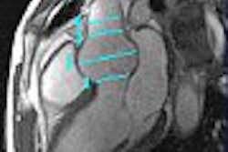
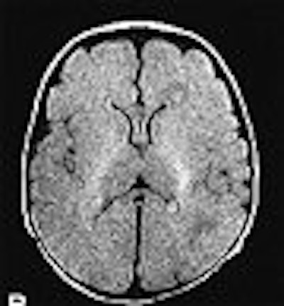
| Images from a normal study of a premature neonate born at 30 weeks' gestation, and imaged 19 days after delivery. Diffusion-weighted image (A); FLAIR image (B). Ultrafast Magnetic Resonance Imaging of the Neonate in a Magnetic Resonance-Compatible Incubator With a Built-In Coil, Whitby EH, Griffiths PD, Lonneker-Lammers T, Srinivasan R, Connolly DJA, Capener D, Paley MNJ, Pediatrics, February 2004, Vol.113, No.2, p.e151. |
No sedation or anesthesia was required. The images were scored for quality by Whitby, who is a neonatal radiologist, as well as by co-authors Paul Griffiths, Ph.D., and Dr. Daniel Connolly, both neuroradiologists.
The group reported good or excellent T1- and T2-weighted images, diffusion-weighted images, and FLAIR images in all seven cases. The neonates remained stable throughout the exams. They found three of the babies to be normal, three with subdural hematoma, and one with germinal matrix hemorrhage.
"T1 data were best obtained from a spin-echo sequence (acquisition time of two minutes and 46 seconds) and T2 weighted from SSFSE (20 seconds)" the group reported.
Future investigations should focus on improving image quality, and, more importantly, making the procedure safer all-around. For now, the neonate is not easily visible from the control room, they cautioned. While the incubator is temperature-controlled, additional monitoring should be done with electrocardiography and MR-compatible pulse oximetry.
Neurodevelopmental handicaps
As the technology for neonate imaging is fine-tuned, pediatric health specialists will gain greater insight into the long-range effects of being born too soon. Even among those who are premature, yet have uncomplicated neonatal courses, serious cognitive and educational problems can still develop.
"The prevalence in this population of major neurodevelopmental handicaps, such as cerebral palsy and mental retardation, ranges from 12% to 23%," wrote Dr. Bradley Peterson from the Columbia College of Physicians & Surgeons and the New York State Psychiatric Institute, both in New York City (Annals of the New York Academy of Sciences, December 2003, Vol. 1008, pp. 219-237).
Structural abnormalities seen with imaging could help determine which pre-term infants would benefit from early, targeted therapeutic intervention. To date, diffusion tensor imaging has indicated the presence of disturbed structural organization of myelinated fibers in premature babies, Peterson pointed out. Metabolically based scans (such as 99mTC-HMPAO SPECT) of preterm infants have shown an increase in blood-flow velocity, which may contribute to hemorrhages in the germinal matrix surrounding the cerebral ventricles, he said.
Peterson's group has performed studies that measured regional brain volumes from the MR scans of 25 prematurely born eight-year-old children. They found that cortical abnormalities were centered on the sensorimotor region of the brain. Also, amygdala and hippocampal volumes were reduced by as much as 30%.
"These findings suggest that volumes of brain regions in preterm neonates may prove to be a useful marker to identify infants at risk for cognitive impairment if they are followed into later childhood," he surmised.
By Shalmali PalAuntMinnie.com staff writer
April 30, 2003
Related Reading
OB ultrasound still tops litigation list, November 12, 2003
Stereotactic radiotherapy effective for pediatric brain tumors, October 23, 2003
Copyright © 2004 AuntMinnie.com