"It is a well-known fact in the radiological community that [chest radiography] has low accuracy," noted Dr. Trond Mogens Aaløkken in an email to AuntMinnie.com. "Why wasn't it replaced by CT a long time ago, just as plain radiographs of the skull, abdomen, and urinary tract have been almost completely replaced by CT or other modalities?"
The research team speculated that this could be attributed to several factors, such as higher radiation dose, greater expense, and more time involved in acquiring CT exams.
Recent studies with low-dose CT have achieved radiation dose ranges of 0.3 to 1.5 mSv, which is an improvement, but it's still considerably higher than the 0.05 mSv dose found in chest radiography. However, new iterative reconstruction techniques are allowing ultralow-dose CT exams to approach the levels of radiography, Aaløkken said.
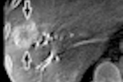
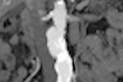
The researchers examined 13 patients with suspected pulmonary lesions, who first received chest radiography and then ultralow-dose CT using an iterative reconstruction protocol (adaptive iterative dose reduction [AIDR] 3D, Toshiba Medical Systems). Image quality was assessed for both radiography and CT by three experienced chest radiologists.
There were a total of 33 findings in the patient group, including 19 nodules. The average sensitivity for all three readers was 18% for radiography and 89% for ultralow-dose CT (p < 0.05). There were 31 false positives in radiography and only two in the CT exams, producing a positive predictive value of 37% for x-ray and 98% for CT. All readers also scored the CT images as having higher quality than x-ray.
But what about radiation dose? The radiography exams had an average radiation dose of 0.05 mSv, while the ultralow-dose CT exams had a dose of just 0.1 mSv -- only twice as much as x-ray. CT exams only took slightly longer to acquire, as well, at seven minutes per study versus five minutes for x-ray.
Ultralow-dose CT demonstrated obvious superiority to radiography in terms of image quality, while radiation dose and acquisition times are comparable, the researchers concluded. The only remaining barrier to increased use of CT in chest imaging is the modality's higher cost, they added.