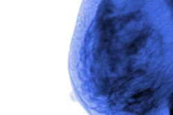
A team led by Dr. Lucas Borges of the University of São Paulo in Brazil developed a denoising tool to address the problem of noise on MBI scans. The tool includes a computer-aided diagnosis feature that searches for and highlights bright regions on the clean image. It also registers the MBI data with digital mammography data and maps coordinates from the MBI exam onto the mammography exam. MBI data can also be fused with corresponding mammography images and displayed in a way similar to PET/CT.
The study included 18 patients. Borges' group found that the algorithm improved the visualization of lesions, and the MBI registration resulted in good matches between MBI and digital mammography images.
The findings suggest that the technique could reduce false positives, Borges and colleagues concluded.
"Denoising may offer the potential to reduce MBI radiation dose and imaging time and increase tumor detectability," they wrote. "By coregistering MBI and digital mammography images, ambiguities between the modalities are reduced, offering the potential to reduce false-positive findings."