While diagnostic tests that measure biomarkers such as carcinoembryonic antigen (CEA) have become important tools in the early detection of disease, such biomarkers are usually only present in low concentrations in blood. Preview research indicates that low-frequency ultrasound compromises the permeability of cell membranes; the Stanford team wanted to see whether such ultrasound waves, applied to potential tumor sites, could release more biomarkers into the bloodstream, thus making tumors easier to detect. The study will be presented by lead researcher Aloma D'Souza, PhD.
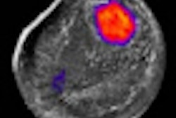
The researchers used a mouse model in which cancer cell lines were exposed to 1-MHz ultrasound of various intensities and durations. Cancer cell lines included colon-LS174T and prostate-LNCaP, which produce biomarkers CEA, carbohydrate antigen 19-9 (CA19-9), and prostate-specific antigen (PSA). Subcutaneous tumors of LS174T in mice (n = 7 per group; n = 28 total) were also sonicated directly over the tumors. In the control group, mice were sonicated on regions without tumors.
The researchers used an enzyme-linked immunoabsorbant assay to detect biomarkers in culture media or murine blood samples. They found that applying 1-MHz ultrasound to LS174T at various intensities intensified the CEA released into the blood.
The researchers also found a substantial increase in PSA when ultrasound was applied to the prostate cancer cell line LNCaP, according to both the intensity and duration of the ultrasound wave.
The technique demonstrates a method for increasing the release of biomarkers in mice by using ultrasound to sonicate cells or tumors, the authors concluded. They believe that the study could demonstrate how medical imaging can become involved in personalized medicine by perturbing lesions and amplifying the release of biomarkers.