"3D MRI captures a larger segment of the carotid artery over a shorter time than 2D MR imaging and allows for more accurate volumetric quantification of intraplaque hemorrhage," Dr. Tishan Maraj, a radiology resident at the University of Toronto, told AuntMinnie.com. "Using a semiautomated method such as this increases consistency of measurements between various readers and sites and would also help monitor and measure progression and regression."
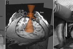
Maraj and colleagues retrospectively evaluated carotid arteries with MRI-detected intraplaque hemorrhage (IPH) using 3D T1-weighted gradient-recalled echo (GRE) MR sequences, which created 2-mm-thick axial reformats for each sequence.
Intraplaque hemorrhage is characterized by high-intensity areas within the vessel wall when compared with the sternocleidomastoid muscle on 3D MRI. Thus, two expert readers identified IPH based on intensities greater than 1.5 times the sternocleidomastoid muscle. They then independently rated randomized images from 10 carotid arteries for the presence of IPH. The goal was to determine if the semiautomated image processing protocol would consistently identify and quantify IPH on 3D MRI with the accuracy of an expert reader.
The results revealed strong agreement between the expert readers for the initial independent assessment, with the receiver operator characteristics (ROC) curve suggesting excellent diagnostic accuracy. The 3D MRI protocol also achieved specificity of 100% and a positive predictive value of 100% for regions of IPH greater than or equal to 1 mm2.
"The immediate clinical benefits are still some time away," Maraj said. "Potentially, it would provide a method for objective identification and measurement of hemorrhage within the carotid artery wall -- a feature that is associated with increased cerebrovascular event occurrence."