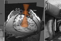
In their study, 185 consecutive patients with histologically confirmed endometrial carcinoma underwent preoperative pelvic 1.5-tesla DCE-MRI. Regions of interest (ROI) for tumors and corresponding normal myometrial ROI were drawn manually on the DCE images at two minutes postcontrast on the slice displaying the largest cross-sectional tumor area.
Low tumor blood flow was among the biomarkers that were significantly associated with high-risk histologic subtypes such as histological grade 3 and nonendometrioid tumors, both of which are associated with reduced survival. The researchers also found a link that suggests tumor hypoxia may lead to more aggressive endometrial cancers.
"Tumor biomarkers from advanced functional MR imaging provide preoperative information that is relevant for clinical stage, histological risk group, and outcome in endometrial cancer patients," study presenter Kristine Fasmer, a medical physicist at Haukeland University Hospital in Norway, told AuntMinnie.com. "The integration of imaging biomarkers in risk-stratified treatment algorithms may ultimately lead to better customized endometrial cancer treatment.