Researchers sought to compare lesion detectability of various MRE imaging techniques in pediatric Crohn's disease patients and evaluate the correlation between detectability grades on each MRE sequence.
"There is no standardized MRE protocol for Crohn's disease patients," said lead study author Dr. Beomseok Sohn, a resident in the department of radiology at Yonsei University in Seoul. "We constructed our routine protocol by enrollment of [MRE] sequences that previously have proven helpful to evaluate Crohn's disease."
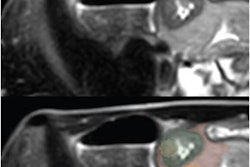
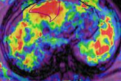
Pediatric patients with confirmed Crohn's disease underwent MRE, including a single-shot fast spin-echo sequence (SSFSE), 2D coronal imaging, diffusion-weighted imaging (DWI), delayed-phase MRE, and arterial dynamic contrast-enhanced MRE.
Lesion detectability was graded on a scale of 0 to 2, with 0 meaning not detected, 1 meaning suspicious, and 2 meaning well-visualized for each MRE sequence for each bowel segment. The study included 35 lesions in 13 patients, who had a mean age of 13.9 years old.
SSFSE fared best, according to the researchers, with a mean detectability grade of 1.94, followed by delayed-phase MRE at 1.74, DWI at 1.69, arterial contrast-enhanced MRE at 1.51, and 2D coronal imaging at 1.11.
The lesion detection rate was 100% on SSFSE sequences, compared with 94% on delayed-phase imaging, 91% on arterial MRE, 89% on DWI, and 80% with 2D coronal imaging.
All MRE sequences showed high lesion detectability for pediatric Crohn's disease patients, although 2D coronal imaging showed the lowest detectability, Sohn and colleagues concluded.