Lead study author Luigi Lepanto, MD, and colleagues reviewed research from the past 10 years dealing with peripheral cystic lesions. Seven articles met selection criteria, with six papers being retrospective studies.
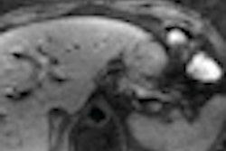
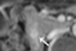
A review of the literature found that overall sensitivity for detecting malignant or premalignant lesions ranged from 65% to 71%, while specificities ranged from 87% to 90%. When specific imaging findings such as thickened septa, lesion size, and wall thickness are considered, sensitivity ranged from 35% to 81%, while specificity ranged from 34% to 75%.
"The ubiquity of medical imaging and the increased sensitivity for the detection of pancreatic lesions will lead to an ever-increasing number of patients being diagnosed with cystic tumors of the pancreas," Lepanto said. "Our systematic review shows that the evidence base for the formulation of practice guidelines is not strong."
The researchers also noted that imaging details were not always fully described in previous studies that were evaluated, and diagnostic criteria are inconsistent from study to study. "In some of the studies," Lepanto added, "overall impression of malignant versus benign is reported, but not the sensitivities and specificities of individual imaging findings. It is difficult to generalize the findings in a coherent way."
To identify helpful imaging and diagnostic guidelines, it will be necessary to design a "pragmatic, prospective study that will allow a better understanding of the evolution of these tumors in a more representative patient population than that studied to date," Lepanto said.