However, in this study, based at Technical University Munich, they concluded that MRI -- at least for the time being -- will remain the standard imaging modality to detect active inflammation in rheumatic diseases.
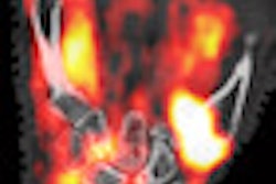
Ten patients (seven females, three males) with a mean age of 52.2 years and clinical suspicion of active inflammation in rheumatoid arthritis were examined with a new optical imaging system from life sciences company Mivenion of Berlin. The device uses the contrast agent indocyanine green in the imaging process.
Three independent radiologists evaluated the degree of inflammation in the metacarpophalangeal, proximal, and distal interphalangeal joints of both hands on a four-point-ordinate scale, ranging from 0 for no inflammation to 3 for severe inflammation.
Those results were compared to scans on a 3-tesla MRI system (Magnetom Verio, Siemens Healthcare, Erlangen, Germany) for a standard of reference.
A total of 280 joints from the 10 patients were evaluated, with 253 (89%) joints showing no inflammation in optical imaging scans. In addition, 27 (11%) joints showed mild, moderate, and severe inflammation.
MR images showed no inflammation in 255 (90%) joints, while 25 (10%) joints showed active inflammation. Using MRI as standard of reference, optical imaging had sensitivity of 84%, specificity of 98%, accuracy of 96%, positive predictive value of 78%, and negative predictive value of 98% for detecting active inflammation in patients with rheumatoid arthritis.
"As we figured, MRI will remain the standard imaging modality to detect active inflammation in rheumatic diseases," said Reinhard Meier, MD, who will present the results at the RSNA meeting. "Furthermore, MRI offers information involving anatomic details, such as bony erosions or tendon status, which is not currently assessable via optical imaging."
He added that the clinical optical imaging device did not detect many inflamed joints, especially very discrete inflammations or inflammation of the flexor tendons, because of excitation and detection only of the dorsal side of the hands.
"However, while the performance of optical imaging up to now is inferior to MRI, it might be nevertheless of substantial added value to clinical examination due to its noninvasiveness, low cost, and easy availability," Meier said.
Meier and colleagues plan to continue their research to improve the sensitivity and specificity of the optical imaging device through image fusion, combining optical imaging with MRI or optical imaging with radiography; automated semiquantitative analysis with a software tool; and correction of systematic signal distortions caused by background noise, individual finger width, and pulse frequencies.