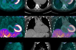
A group led by Dr. Johannes Grueneisen from Essen University Hospital in Germany reviewed 44 consecutive patients with histopathologically confirmed cervical cancer who underwent whole-body scanning with the hybrid modality before therapy. The protocol included dedicated imaging of the female pelvis, followed by assessment by two radiologists in separate reads of datasets produced by PET/MRI and MRI alone. The researchers sought to determine any spread of primary tumors of the uterine cervix, in addition to detecting nodal and distant metastases.
MRI and PET/MRI correctly detected 43 (8%) of the 44 primary cervical tumors. The lone exception was a case of stage IA cancer, which was not found by either imaging technique. In addition, MRI and PET/MRI allowed for a correct determination of tumor stage in 38 (86%) of the 44 patients. Lymph node metastases were present in 19 (43%) of the 44 patients.
PET/MRI achieved greater sensitivity, specificity, and diagnostic accuracy than MRI for identifying nodal-positive patients. In three patients, distant metastases were present and could be detected in both imaging modalities.
The findings demonstrate the usefulness of FDG-PET as a "valuable additive to MR imaging for more accurate nodal staging of patients with cervical cancer," Grueneisen and colleagues concluded. However, FDG-PET did not add any additional information to MRI for identifying the spread of local tumors.
The combination of MRI and simultaneous PET advances the evaluation of uterine cervical cancer and could become a "valuable alternative/adjunct for the clinical workup in a pretreatment setting," they wrote.