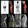
"Based on the high soft-tissue contrast capabilities of the MR component, especially small lesions of the liver can be better identified than on CT," noted senior study author Dr. Patrick Veit-Haibach from the department of medical imaging at University Hospital Zurich. "The benefits are a potentially better diagnosis by identifying more lesions. There is also the benefit of having PET/CT and MRI together, coregistered and being done on the same appointment."
The downside of the study is that no difference in lesion detection or localization was seen, so PET/MRI had no benefit in cancer staging, according to the researchers.
In the study, eight women and seven men (mean age, 52.3 years) with various oncological diseases were referred for a clinical PET/CT scan. An additional PET/MRI was performed within a trimodality PET/CT-MRI system with ultrafast GRE and SE sequences and body-array coils covering the entire abdomen.
All lesions (lymph node metastases/organ metastases) were compared for detection rate and localization, lesion size, and conspicuity. In all, the researchers evaluated 37 PET-positive lesions (29 abdominal lesions and eight pelvic lesions).
They found no difference between modalities for lesion detection and localization. However, there was a statistically significant difference between the mean size of the lesions (17.2 mm for CT; 27.8 mm for MRI), as well as for overall lesion conspicuity (mean score of 2.35 for PET/CT; 2.85 for PET/MRI).
"We are beginning to optimize our PET/MRI protocols for abdominopelvic cancer, and this study with nonenhanced MR imaging can be seen as a basic work," added study co-author Dr. Diethard Schmidt. "Besides contrast studies, there are many possibilities to improve further PET/MRI protocols. The challenge will be to develop fast and robust PET/MRI protocols to manage the daily practice in oncologic imaging."
In the future, there is great potential for MRI with additional biomarker functions such as diffusion-weighted sequences, diffusion tensor imaging, and other imaging technologies to combine with PET to improve diagnosis of and therapy for cancer, Schmidt said.
