The study, from the University of Leipzig in Germany, noted that with aging, the radial structure of the cartilage becomes more isotropic. T2-weighted 7-tesla MRI data and a simplified structure model would enable radiologists to derive quantitative parameters of the collagen fiber arrangement and to estimate the thickness of the radial zone in adult cartilage. The method could therefore be used to noninvasively determine the "biologic age" of the cartilage.
Study co-author Nikita Garnov, PhD, noted that the aging process leads to degenerative changes in cartilage tissue, which can cause arthritis. Therefore, a noninvasive diagnostic technique is essential for early assessment to help to slow that degenerative process.
"MRI holds great promise in the early diagnostics of arthritis, in particular because of its noninvasiveness," said Garnov, who will present the study results. "Routine MRI, however, is practically not suited for this task due to the low spatial resolution."
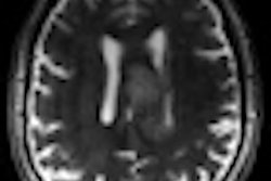
Researchers evaluated the right knees of 10 healthy volunteers between the ages of 21 and 30 years on a 7-tesla whole-body MRI scanner. Sagittal images of the lateral condyles were acquired with an eight-channel phased-array coil using a T2-weighted multiecho spin-echo sequence.
"The present study relies on both 7-tesla MR imaging and an advanced technique for subsequent analysis of the cartilage ultrastructure," Garnov explained. "Highly resolved T2-weighted images of the cartilage are then analyzed using an angle-dependent model to determine the structural fiber arrangement. In particular, we derive the relative thickness of the radial layer, which can be used as an indicator for structural order and state of health, or biological age, of the cartilage tissue, because the radial structures are expected to become less organized with aging."
The researchers found that high-resolution 7-tesla MR images of the knee cartilage had a sufficient signal-to-noise ratio for subsequent quantitative analysis. The MRI scans also revealed that cartilage thickness was between 2.6 mm and 3.8 mm in the femur and between 3.2 mm and 5.2 mm in the tibia.
"Due to the higher signal-to-noise ratio at 7 tesla at relatively small voxel sizes, ultrahigh-field MRI is a promising tool to investigate joint cartilage on a small-length scale, thickness less than 5 mm, in vivo," Garnov added.
Currently, Garnov and colleagues are planning measurements on the knees of volunteers between the ages of 50 and 60 years to evaluate potential differences in the cartilage ultrastructure between younger and older people.
They also are looking to evaluate whether the main findings at 7 tesla MRI already can be obtained in whole-body scanners at lower field strengths, in particular 3-tesla magnets, which are more commonly available and would also have the advantage of allowing actual patient studies.
"At 7 tesla, ethical concerns of patient studies are a major issue and approval is generally still difficult," Garnov said. "In addition, we plan to investigate the behavior of the collagen structure under in-situ load bearing of the knee joint in 1.5-tesla or 3-tesla MRI."