The researchers have proposed a novel method for automatically segmenting functional and anatomical images that is ready to be used in clinical routines, said presenter Ulas Bagci, PhD, of the NIH.
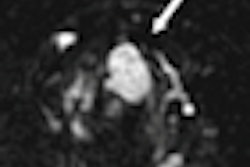
Their computational platform can perform these segmentations within seconds and perform coevaluation based on the segmentation results. Effectiveness of the method was validated using PET phantoms, clinical PET studies, CT exams, PET/CT studies, and PET/MRI images, Bagci said.
"Our method is not limited to PET/MRI but can be used in PET/CT as well," he said.
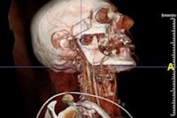
Accurately assessing radiotracer uptake from PET and extracting corresponding anatomical tissue boundaries from MRI simultaneously is an important application in computational radiology, radiotherapy planning, surgery planning, and other diagnostic tasks, Bagci said.
"Our study is important because methods to improve segmentation from fused imaging modalities increase the accuracy and efficiency of quantitatively measuring disease and response to therapy by simultaneously using functional molecular data from PET and anatomical data from MRI or CT," Bagci said. "The presented automated computational tool can be employed for routine use in clinics, accurately identifying intricate boundaries between disease and healthy tissues using joint knowledge from both anatomical and functional information."