A group from the Mayo Clinic in Rochester, MN, aimed to develop and validate a faster way of estimating patient attenuation, with scout images as one possibility, for example.
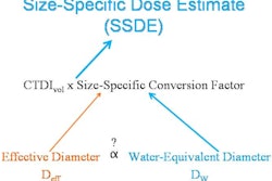
"To make use of ... SSDE recently published by the American Association of Physicists in Medicine, a robust automated method is needed to automatically quantify patient size," co-author Cynthia McCollough, PhD, told AuntMinnie.com.
The researchers described attenuation in terms of a water cylinder having the equivalent absorbed dose, calculating the cross-sectional area using CT images and incorporating the CT value and pixel numbers. They weighted the dependence of the cross-sectional area on attenuation using both linear and quadratic dependence methods. Radiation dose to the phantoms was calculated using validated Monte Carlo methods and clinical scanning parameters.
The Monte Carlo simulations showed that the cross-sectional area in a thorax phantom varies with object attenuation in a mostly linear fashion. Using only geometrical information -- as would be the case in physical measurements -- underestimated the dose by about 10%, while attenuation information obtained with the formula erred by less than 2% for adult phantoms and less than 6% for pediatric phantoms.
"Patient size can be easily and reproducibly quantified in terms of what matters most for dose estimation, which is not dimension or weight, but x-ray attenuation," McCollough said.