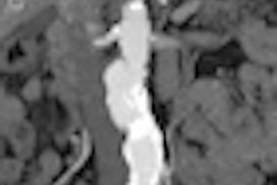
Lymphadenopathy is often an indicator of other underlying disease, and automatic detection of these enlarged lymph nodes could reduce errors and increase interpretative efficiency compared with manual diagnosis. However, most CAD techniques still suffer from high-false positive rates, said lead author Jiamin Liu, PhD, a staff scientist at the U.S. National Institutes of Health.
Because anatomical relationships such as the locations of candidate lymph nodes relative to the major organs could provide important information for lymph node detection, the researchers applied the detection of the liver and spleen for CAD of lymphadenopathy.
Not only did the new technique yield higher sensitivity over CAD without organ detection (90% versus 75% at 10 false positives per patient), it also cut false positives by 50% (10 versus 20 at 90% sensitivity), he said.
"Our fully automatic technique shows promise for accurate detection of lymphadenopathy in contrast-enhanced abdominal CT scans," Liu said. "Segmented organs [liver and spleen] can further improve the CAD performance by markedly improving sensitivity and reducing false positives."