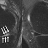
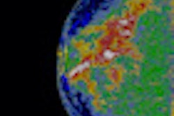
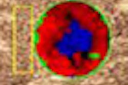
Shoulder instability is a complex disorder that requires the combination of multiple imaging modalities to best define anatomical-pathological changes. A significant amount of bone loss from the anteroinferior glenoid has also been shown to be a predisposing factor for recurrent instability and a negative prognostic factor for arthroscopic capsular repair, said presenter Massimo De Filippo, MD, of the University of Parma.
Based on the need to identify and quantify glenoid bone loss preoperatively, the researchers sought to determine the accuracy of CT curved MPR in quantifying glenoid bone loss in patients with monolateral recurrent shoulder dislocation.
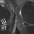
The researchers found that curved MPR CT and arthroscopy findings agreed in all 12 patients with bony Bankart lesions. They also determined that glenoid bone loss was accurately calculated by curved MPR CT in 30 of 32 patients (94% sensitivity). In addition, the absence of bone loss was correctly identified in both patients without bone loss (100% specificity).


