Radiographics 1992 Sep;12(5):1013-1030. From the archives of the AFIP. Mediastinal germ cell tumors: radiologic and pathologic correlation.
Rosado-de-Christenson ML, Templeton PA, Moran CA
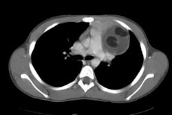
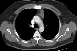
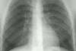
Germ cell tumors occur most frequently in the gonad but can rarely occur in extragonadal locations, usually in or near the midline. The most common extragonadal site of primary germ cell tumors is the anterior mediastinum. The most common histologic type of mediastinal germ cell tumor is mature teratoma, which is typically asymptomatic and incidentally discovered. Radiographically, these tumors appear as rounded, often lobulated masses; calcification may be seen. Imaging studies of mature teratoma frequently demonstrate cystic components and may demonstrate fat or calcium. Malignant germ cell tumors usually occur as large masses in symptomatic young male patients. Seminomas are typically of homogeneous soft-tissue attenuation, and nonseminomatous malignant germ cell tumors are typically of heterogeneous attenuation on computed tomographic scans. Therapy varies according to cell type and may include surgery, radiation therapy, or chemotherapy. Prognosis is excellent for patients with mature teratoma, good for patients with pure seminoma, and poor for patients with nonseminomatous malignant germ cell tumors and mixed germ cell tumors.
PMID: 1326777, MUID: 92410031