Intralobar bronchopulmonary sequestration
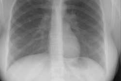
The patient below presented for evaluation of an abnormal CXR which demonstrated a left retrocardiac mass. (Click image to enlarge)

CT scan revealed a soft tissue mass in the left posterior-medial lung which had a branching tubular appearance. Some associated para-emphysematous changes were noted. (Click images to enlarge)


An arteriogram was performed and reveal a systemic arterial supply to the mass with pulmonary venous drainage. The mass was resected and determined to be an intralobar bronchopulmonary sequestration.