CHICAGO - CT images are helpful for planning a complicated surgery, but physicians say that being able to hold a patient's skull in their hands makes surgical planning for face transplantation much easier. And this can be accomplished with 3D printing technology, researchers explained at the RSNA 2014 meeting.
"This is a complex surgery, and its success is dependent on surgical planning," said Dr. Frank Rybicki, director of the Applied Imaging Science Laboratory at Brigham and Women's Hospital. "Our study demonstrated that if you use this model and hold the skull in your hand, there is no better way to plan the procedure."
When a face transplant candidate is evaluated, the researchers use CT imaging with 3D visualization to create life-sized structures. CT images of the skull are segmented and processed using customized software and files are delivered to a 3D printer (Objet, Stratasys; Materialise).
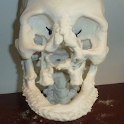
 3D-printed facial model. Image courtesy of RSNA.
"It takes a few hours to produce the skull and facial features," Rybicki said, illustrating for AuntMinnie.com how the pieces fit together, giving doctors the ability to better understand the patient's condition. At the press conference at RSNA, Rybicki showed how the 3D-printed skull of face transplant recipient Carmen Tarleton conformed to the soft-tissue 3D-printed polymer.
"This is an exploratory way of evaluating soft tissues," he said. The research team is giving hands-on demonstrations of the technology at the meeting, he added.
"We are demonstrating that 3D printing is feasible in the surgical planning and follow-up of soft tissue changed in the healing process," said Dr. Amir Imanzadeh, a research fellow at Brigham and Women's and member of the face transplant team.
Sometimes the transplant recipient's facial bones need to be modified prior to the surgery, Imanzadeh said. The 3D model helps with this preparation, so that the actual surgery proceeds more smoothly.
 Dr. Frank Rybicki with the 3D-printed skull and face.
"If there are absent or missing bony structures needed for reconstruction, we can make modifications based on the 3D printed model prior to the actual transplantation, instead of taking the time to do alterations during ischemia time [when blood flow is stopped]," Rybicki said. "The ability to work with the model gives you an unprecedented level of reassurance and confidence in the procedure."
The models displayed at the conference and during the educational sessions were 3D printed directly from standard CT images, said Dr. Catherine Phillips, a resident at Brigham and Women's.
"This gives a whole different tactile element to our presurgical planning," she said.
Dr. Donald Annino, an assistant professor of surgery at Harvard Medical School, told AuntMinnie.com that from the point of view of the surgeon, "the 3D printed skull is extremely important in planning patients with skull deficiencies."
The Brigham team has participated in several face transplants performed in the U.S. The researchers said that 30 transplants have been performed worldwide.
Face transplant recipient Carmen Tarleton, who attended the press conference, said she experienced third- and fourth-degree burns after her ex-husband poured industrial lye on her face. In addition to reducing her pain, the face transplantation gave her new lips and eyelids and helped restore partial vision in one of her eyes.
"This whole experience of having a face transplant has been life-changing for me," she said. "The pain, especially in my neck, was removed instantaneously with the face transplant."
She said she appreciates the vast number of people who helped develop the technology, in and out of the medical field, "that has helped me so greatly."