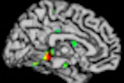
Changes in tumor enhancement on postgadolinium T1-weighted MRI scans are highly predictive of which patients with recurrent glioblastoma will benefit from second-line therapy that includes the antiangiogenic drug bevacizumab, according to a study presented last week at the RSNA 2012 conference.
Determining whether a patient's tumor is responding to a therapeutic regimen with bevacizumab has presented challenges in MRI interpretation due to the drug's potential for decreasing tumor enhancement independent of decreasing tumor burden.
The findings were presented by Dr. Jerrold Boxerman, PhD, assistant professor at the Warren Alpert Medical School of Brown University, vice chair for brain cancer research of the American College of Radiology Imaging Network (ACRIN) head/neck/neuro committee, and principal investigator for the ACRIN 6677 trial.
In a review of 123 cases, two radiologists measured 2D and 3D enhancement on postgadolinium T1-weighted MR images and 3D fluid-attenuated inversion-recovery (FLAIR) hyperintensity images. A third neuroradiologist reviewed any interpretation discrepancies.
The median overall survival of patients whose tumors had progressed after two cycles and four cycles of treatment (at eight and 16 weeks, respectively) was significantly less than that of patients whose tumors did not progress on the 2D T1 images and 3D T1 images, but not on the FLAIR images.
"Our study results demonstrate that even though tumor enhancement may diminish with the use of bevacizumab, a finding of progressive enhancement on the T1-weighted images highly correlates with worse patient outcome," Boxerman said in a statement about the study. "Knowing that a patient is not benefiting from a treatment could permit a timely switch to a clinical trial evaluating an alternate therapeutic strategy."