CHICAGO - Use of diffusion-tensor MRI (DTI-MRI) could help detect subtle injuries that can last from hours to months in patients with mild traumatic brain injury, according to a study presented on Monday at the RSNA annual meeting.
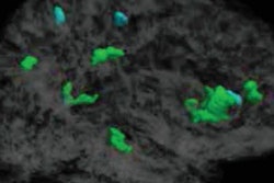
With DTI, doctors can measure the uniformity of water flow, or fractional anisotropy, throughout the brain, said Dr. Michael Lipton, associate director of the Gruss Magnetic Resonance Research Center at the Albert Einstein College of Medicine.
Low fractional anisotropy indicates regions of axonal injury, whereas abnormally high fractional anisotropy may indicate areas where the brain is attempting to compensate for the injury, he said.
To determine the long-term impact of concussion, Lipton and colleagues recruited 25 patients from emergency room visits, most of whom had suffered injury in assaults or motor-vehicle accidents. They were matched with 40 control subjects who were rigorously interviewed to determine that they had no history of concussion.
Lipton said in a press briefing that about 30% of patients with mild traumatic brain injury have long-standing postconcussion symptoms. Current determination of whether recovery is complete is usually based on physical functioning. However, use of DTI may be able to determine how well that recovery is going, he suggested.
"Just because someone looks like they're back to normal doesn't mean their brain has compensated for the injury," he said.
In the study, Lipton and colleagues performed DTI-MRI on all patients within two weeks of their injury, evaluating whether fractional anisotropy was uniform as in healthy brains or disturbed, indicating concussion. A year after their brain injury, the patients also completed questionnaires to assess postconcussive symptoms and health-related quality of life.
Patients exhibiting higher levels of fractional anisotropy showed greater improvement in concussive symptoms (p = 0.01). They also performed better in quality-of-life measures of mobility control (p = 0.024) and psychological functioning (p = 0.007).
The results indicate that concussion is an injury that may evolve over a period of time, Linton said. He acknowledged that further studies are needed to determine the role of DTI and fractional anisotropy in mild traumatic brain injury, but it eventually might be useful in predicting outcomes for concussion patients, as well as for guiding new treatment approaches.
"The science around this potential biomarker in concussion is just developing," said Dr. David Hovsepian, professor of radiology at Stanford University Medical Center, who moderated the press briefing. "How this description of what happens in the brain fits into clinical practice is not yet known."